Comparative Study
. 2004 Jul;39(3):155-66.
doi: 10.1002/gene.20036.

Folate modulates Hox gene-controlled skeletal phenotypes

Affiliations
Comparative Study

Folate modulates Hox gene-controlled skeletal phenotypes

Claudia Kappen et al. Genesis. 2004 Jul.

Abstract

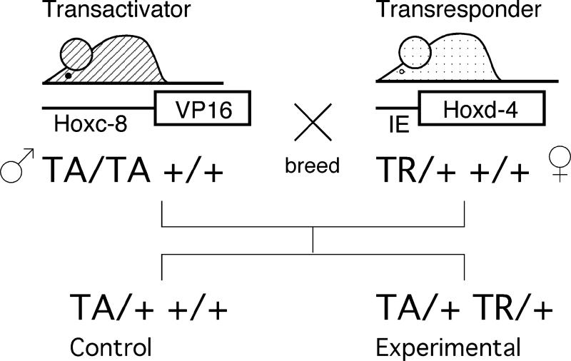
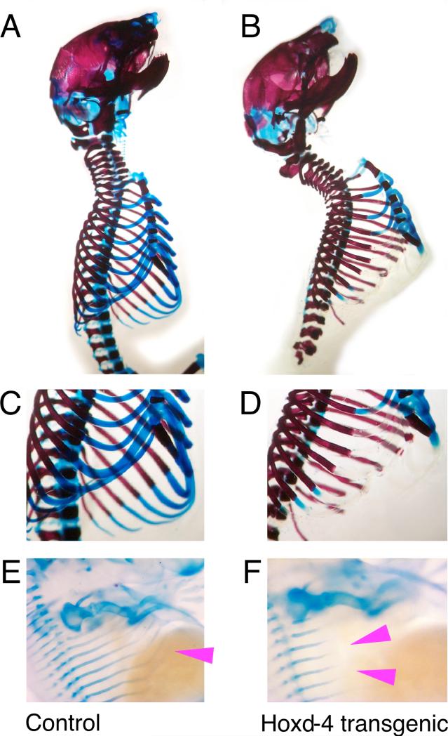
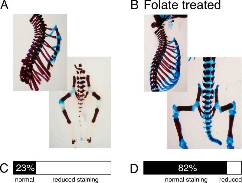
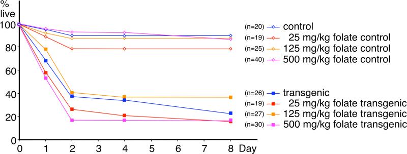
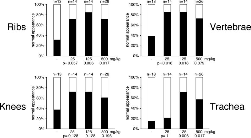
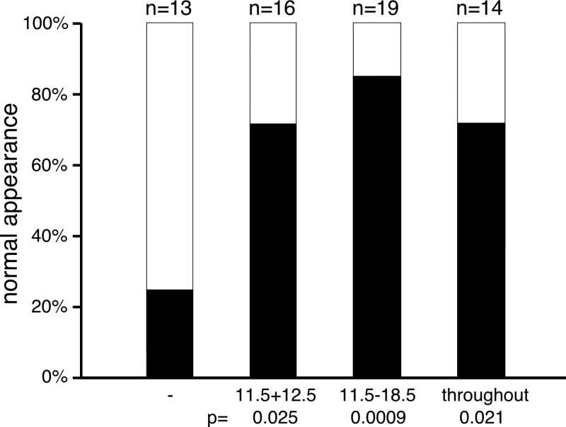
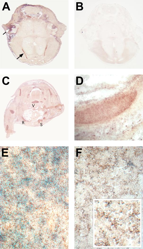
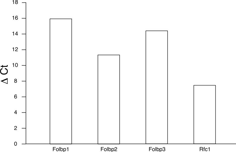
Hox genes are well-known regulators of pattern formation and cell differentiation in the developing vertebrate skeleton. Although skeletal variations are not uncommon in humans few mutations in human HOX genes have been described. If such mutations are compatible with life, there may be physiological modifiers for the manifestation of Hox gene-controlled phenotypes, masking underlying mutations. Here we present evidence that the essential nutrient folate modulates genetically induced skeletal defects in Hoxd4 transgenic mice. We also show that chondrocytes require folate for growth and differentiation and that they express folate transport genes, providing evidence for a direct effect of folate on skeletal cells. To our knowledge, this is the first report of nutritional influence on Hox gene-controlled phenotypes, and implicates gene-environment interactions as important modifiers of Hox gene function. Taken together, our results demonstrate a beneficial effect of folate on skeletal development that may also be relevant to disorders and variations of the human skeleton.


Figures

Figure 1
Figure 1. Generation of Hoxd4 transgene expressing mice in the VP16 binary transgenic system
Two independent parental strains carry the transactivator (TA) and transresponder (TR) transgenes, respectively. Animals from both parental strains are normal; activation of the TR transgene is achieved only in simultaneous presence of the TA transgene. Experimental and control animals are generated within the same female, providing for comparable nutritional exposure of controls and Hoxd4 transgenic animals.
Figure 2
Figure 2. Cartilage defects in Hoxd4 transgenic mice
Preparations of skeletons stained with Alizarin Red for bone and Alcian Blue for cartilage. Rib cages of (A,C,E) control and (B,D,F) Hoxd4 transgenic newborn mice (genotype TA/+ TR/+). (A,B) Staining for Alcian blue is absent or reduced in ribs (arrowhead) of the Hoxd4 transgenic animal (B,D). (C,D) Close-ups of rib cages caudal from the third rib in newborn skeletons. Embryos were isolated at gestational day 13.5 and stained for Alcian Blue. (E) Magnification of a control embryo showing the rib cartilage anlagen (arrowhead); their extension is reduced upon expression of the Hoxd4 transgene (F), indicating delayed cartilage formation and maturation (compare position of arrowheads). This embryo had the genotype TA/TA TR/+.
Figure 3
Figure 3. Folate supplementation restores Alcian Blue staining in transgenic cartilage
(A) Skeleton of a Hoxd4 transgenic newborn mouse. (B) Skeleton of a Hoxd4 transgenic raised in a mother who received folate daily throughout the pregnancy at 25 mg/kg body weight. Fraction of skeletons with obvious staining defects in (C) unsupplemented (n=13) and (D) folate-supplemented (n=16) animals. Represented in (D) are results for animals supplemented with 25 mg folinic acid/kg body weight from gestational day 11.5 to 18.5, the group for which the best improvement in most skeletons was noted (p=0.0009). If results for all supplemented animals (n=89; summarizing all regimens) are compared to unsupplemented Hoxd4 transgenic animals (n=56; including all bedding conditions), the fraction of skeletons deficient in cartilage staining was reduced from 59% to 34% (p=0.0035). Alcian Blue staining in ribs and vertebral column is restored by folate supplementation, and rigidity of the skeleton is improved.
Figure 4
Figure 4. Effect of folate supplementation on survival of Hoxd4 transgenic mice
The proportion of live mice declines rapidly after birth for Hoxd4 transgenic newborns (closed symbols), but not for controls (open symbols). The attrition rate for transgenic mice is independent of the dose of folinic acid (different colors), and there are no significant differences between the survival curves for the different transgenic groups (numbers of individuals in brackets), or between control groups.
Figure 5
Figure 5. Differential effect of folate in different cartilage structures
Restoration of Alcian Blue staining in different cartilage structures was assessed independently by two technicians for each skeleton without prior knowledge of genotype. Filled bars represent normal appearance of cartilage; open bars represent unstained or very weakly stained structure. All p-values are calculated relative to the untreated control. Rib (A) and vertebral (B) cartilages were restored by folate supplementation, while knee cartilage (C) did not respond in statistically significant fashion (compare p-values) to folate supplementation. Increased Alcian Blue staining in trachea (D) was only observed at higher doses of folate. Structural rigidity of the vertebral column was found in 23% of untreated controls, but restored in 92.9% of animals with folate supplementation of 25mg/kg (p=0.00034), in 85.7% with 125mg/kg (p=0.0018), and in 65.4% with the 500mg/kg dose (p=0.019). Vertebral column and ribs showed the best improvement of cartilage production and staining with folate supplementation.
Figure 6
Figure 6. Temporal window for cartilage rescue by folate supplementation
Supplementation with folinic acid at 25mg/kg was performed either throughout the pregnancy (see Methods), or restricted the second half of the pregnancy, or gestational days 11.5 and 12.5. Restored cartilage in Hoxd4 transgenic skeletons was observed even with only two days of supplementation. Improvement for ribs and vertebrae was statistically significant (p=0.000025 for E11.5-18.5; p=0.0027 for E11.5/12.5); the longer supplementation lead to some improvement for knees (p=0.005 for E11.5-18.5; p=0.14 for E11.5/12.5) but little for trachea (p=0.062 for E11.5-18.5; p=0.66 for E11.5/12.5). Rigidity of the vertebral column was significantly restored with both treatment regimens (p=0.0009 for E11.5-18.5; p=0.00001 for E11.5/12.5). Supplementation of pregnancies with folate prior to overt skeletogenesis (E11.5/12.5) restores production and staining of normal cartilage in Hoxd4 transgenic mice.
Figure 7
Figure 7. Expression of Folbp2 in developing cartilage
In situ hybridization to sections from a mouse embryo at 15.5 days of development (A-D) and to cultured primary chondrocytes (F). A, Transverse section through the head reveals strong signal for Folbp2 in the choroid plexus (closed arrow) and also in developing cartilage (open arrow points to ear cartilage as an example). This signal is specific for the Folbp2 probe; a control section from the same embryo processed identically but without probe (B) does not reveal any AP activity. (C) Folbp2 expression is also found in limb and trunk cartilage, such as R: rib, S: sternum, and V: vertebral center. (D) Close-up with signal of rib cartilage. All sections are oriented with dorsal to the top. (E) Primary neonatal rib chondrocytes after 4 days in high-density culture. Cells were stained with Nuclear Fast Red to reveal nuclei, and with Alcian Blue to reveal production of extracellular cartilage matrix. (F) In situ hybridization for Folbp2 to chondrocytes cultured as shown in E. Folbp2 signal is evident in cultured chondrocytes with smaller, proliferating cells expressing Folbp2 at apparently higher levels than mature hypertrophic cells (inset).
Figure 8
Figure 8. Chondrocytes express genes encoding folate transport molecules
Quantitative RT-PCR was performed in triplicate on pooled chondrocytes from normal FVB neonates. GAPDH (Glyceraldehydephosphate Dehydrogenase) expression was used as the standard, and results are expressed as the difference in threshold cycle number for detection of each gene relative to the threshold cycle number for GAPDH in the same sample (CtGENE - CtGAPDH = Δ Ct). Higher numbers indicate that more cycles are needed to detect expression, hence reflecting lower expression levels. The major folate transporters expressed in primary chondrocytes are Folbp2 and Rfc1.
Figure 9
Figure 9. Chondrocytes require folate for growth and differentiation
Primary rib chondrocytes from neonatal mice were placed at high density into culture in folate-containing (FC) or folate-free medium (FF), with or without serum (S). Green: folate and serum, turquoise: no folate but serum, purple: folate but no serum, orange: neither folate nor serum, red: folate and serum and methotrexate (MTX). Serum is able to provide folate (and other nutrients) to folate-depleted medium; without serum, cells in folate-containing medium initially divide, but are unable sustain proliferation (purple) or to differentiate to hypertrophy (not shown). Without folate (orange), or with inhibited folate metabolism (methotrexate, red), chondrocytes are unable to grow and differentiate.

Similar articles

Cited by

References

    1. Akarsu AN, Stoilov I, Yilmaz E, Sayli BS, Sarfarazi M. Genomic structure of HOXD13 gene: a nine polyalanine duplication causes synpolydactyly in two unrelated families. Hum Mol Genet. 1996;5:945–952. - PubMed
    1. Antony AC. Folate Receptors. Annu Rev Nutr. 1996;16:501–521. - PubMed
    1. Barbera JP, Rodriguez TA, Greene ND, Weninger WJ, Simeone A, Copp AJ, Beddington RS, Dunwoodie S. Folic acid prevents exencephaly in Cited2 deficient mice. Hum Mol Genet. 2002;11:283–293. - PubMed
    1. Berry RJ, Li Z. Folic acid alone prevents neural tube defects: evidence from the China study. Epidemiology. 2002;13:114–116. - PubMed
    1. Bird A. DNA methylation patterns and epigenetic memory. Genes Dev. 2002;16:6–21. - PubMed

Publication types

LinkOut - more resources