

Diagnostic accuracy of ultrasonography compared to unenhanced CT for stone and obstruction in patients with renal failure
- PMID: 15283870
- PMCID: PMC514525
- DOI: 10.1186/1471-2342-4-2
Diagnostic accuracy of ultrasonography compared to unenhanced CT for stone and obstruction in patients with renal failure
Abstract
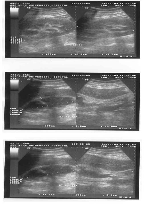
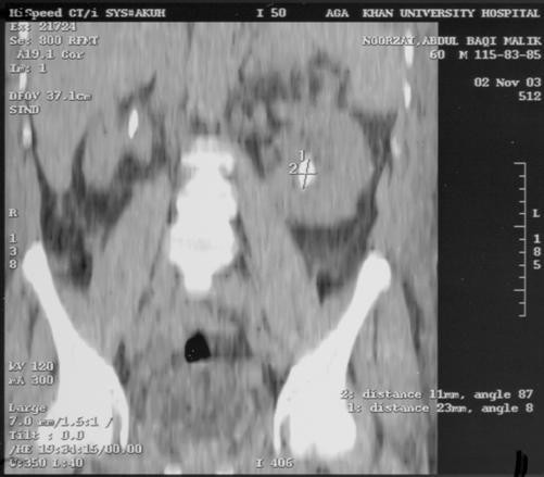
BACKGROUND: To determine accuracy of ultrasound (US) kidney, ureter and bladder (KUB) compared to un-enhanced helical CT (UHCT) in patients with renal failure in the diagnosis of stone and obstruction. METHODS: This is a case controlled study conducted in the period from June 2000 to July 2003 at a university hospital. All patients had both US and UHCT scan. Patients with serum creatinine >/= 1.8 mg/dl were included in the study. Only direct visualization of stone was considered as confirmatory. In both the studies, UHCT and US, presence of stone and obstruction were noted. The relevant biochemicals, radiological and clinical records of all the patients were analyzed. Data was analyzed using commercially available software. RESULTS: During the period of study 864 patients had UHCT for evaluation of the urinary tract in patients presenting with flank pain. Out of these 34 patients had both UHCT and US done within a span of one day and had serum creatinine of >/=1.8 mg/dl. Mean age was 48 +/-15.8 years and 59% of patients were males. UHCT identified renal stones in 21 (62%), whereas 17 of these were identified on US, with a sensitivity of 81%. Of the four patients with renal stones missed on US, three were identified on plain x-ray; the mean size of stones missed was 6.3 mm. Of the 22 (65%) patients with ureteric stone on UHCT, US could only identify 10; a further 7 were identified on x-ray KUB, giving a sensitivity of 45% (US alone) and 77% (US with x-ray KUB). CONCLUSIONS: US is sensitive and specific for renal stones, 81% and 100% and for hydronephrosis, 93% and 100%, respectively. Its sensitivity to pick ureteric stone (46%) and to identify hydroureter (50%) is low. Addition of x-ray KUB abdomen increases the sensitivity for ureteric stones to 77%.
Figures
References
-
- Sudah M, Vanninen RL, Partannen K, et al. patients with acute flank pain: comparison of MR urography with un enhanced helical CT. Radiology. 2002;223:98–105. - PubMed
-
- Smith RC, Rosenfield AT. Acute flank pain: comparison of non-contrast enhanced CT and intravenous pyelography. Radiology. 1995;194:789–794. - PubMed
-
- Sommer FG, Jeffrey RB, Rubin GD. Detection of ureteral calculi in patients with suspected renal colic: value of reformatted non contrast helical CT. AJR Am J Roentgenol. 1995;165:509–13. - PubMed
-
- Guest AR, Cohan RH, Korobkin M, et al. Assessment of clinical utility of the rim and comet-tail signs in the differentiating ureteral stones from phleboliths. AJR Am J Roentgenol. 2001;177:1285–91. - PubMed
LinkOut - more resources
Full Text Sources
