

Acute nociceptive somatic stimulus sensitizes neurones in the spinal cord to colonic distension in the rat
- PMID: 15284340
- PMCID: PMC1665216
- DOI: 10.1113/jphysiol.2004.069070
Acute nociceptive somatic stimulus sensitizes neurones in the spinal cord to colonic distension in the rat
Abstract
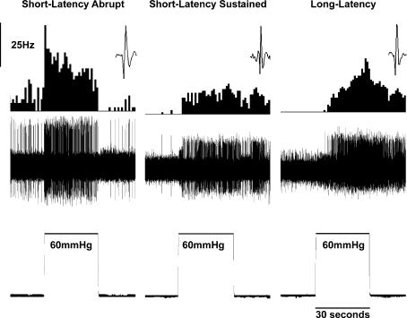
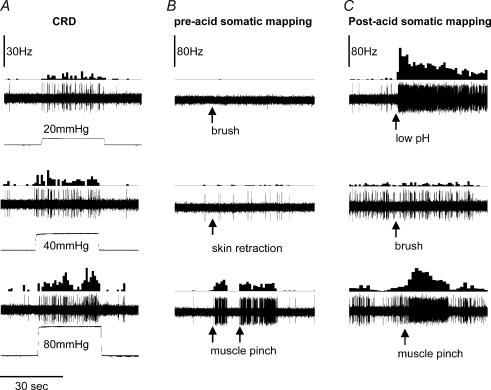
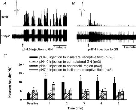
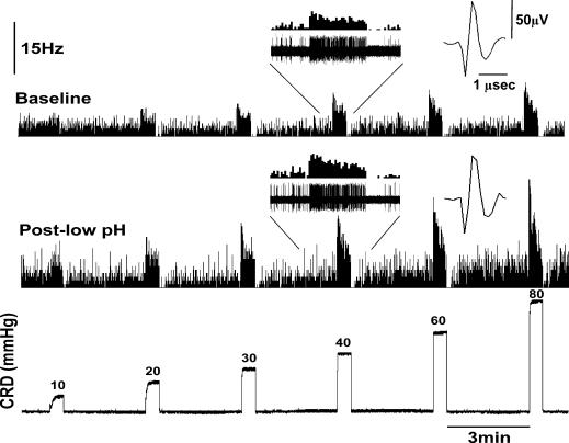
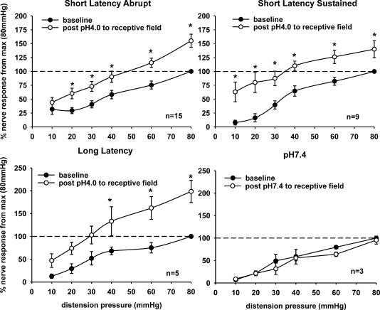
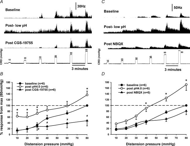
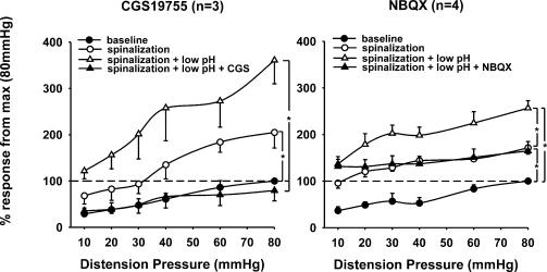
The common co-existence of fibromyalgia and chronic abdominal pain could be due to sensitization of spinal neurones (SNs), as a result of viscero-somatic convergence. The objective of this study is to explore the influence of acute nociceptive somatic stimulation in the form of acid injections, into the ipsilateral somatic receptive field of neurones responsive to colorectal distension (CRD), and the potential role of ionotropic glutamate receptors on sensitization. Action potentials of CRD-sensitive SNs were recorded extracellularly from the lumbar (L(2)-L(5)) spinal cord. Stimulus-response functions (SRFs) to graded CRD (10-80 mmHg, 30 s) were constructed before and 30 min after ipsilateral injection of low pH (4.0, 100 microl) saline into the somatic receptive fields. In some experiments, cervical (C(1)-C(2)) spinalization was performed to eliminate supraspinal influence. The selective NMDA receptor antagonist CGS 19755 and AMPA receptor antagonist NBQX were injected (25 micromol kg(-1), i.v.) to examine their influence on sensitization. Three types of neurones were characterized as short-latency abrupt (SLA, n = 24), short latency sustained (SLS, n = 12), and long-latency (LL, n = 6) to CRD. Ipsilateral injection of low pH (4.0) in the somatic receptive field, but not the contralateral gastrocnemius (GN) or front leg muscles, sensitized responses of these neurones to CRD. Spinalization had no influence on the development of low pH-induced sensitization. Both CGS 19755 and NBQX significantly attenuated the sensitized response to CRD in intact and spinalized animals. Acute nociceptive somatic stimulus sensitizes CRD-sensitive SNs receiving viscero-somatic convergence. The sensitization occurs at the spinal level and is independent of supraspinal influence. Ionotropic glutamate receptors in the spinal cord are involved in sensitization.
Figures
Similar articles
-
Modulation of spinal visceral nociceptive transmission by NMDA receptor activation in the rat.J Neurophysiol. 1996 Jun;75(6):2344-53. doi: 10.1152/jn.1996.75.6.2344. J Neurophysiol. 1996. PMID: 8793747
-
NMDA receptor mediates chronic visceral pain induced by neonatal noxious somatic stimulation.Eur J Pharmacol. 2014 Dec 5;744:28-35. doi: 10.1016/j.ejphar.2014.09.034. Epub 2014 Sep 30. Eur J Pharmacol. 2014. PMID: 25281204 Free PMC article.
-
Differential effects of spinal CNQX on two populations of dorsal horn neurons responding to colorectal distension in the rat.Pain. 2002 Sep;99(1-2):217-22. doi: 10.1016/s0304-3959(02)00106-9. Pain. 2002. PMID: 12237199
-
Responses of rat spinal neurones to natural and electrical stimulation of colonic afferents: effect of inflammation.Brain Res. 2000 Jun 2;866(1-2):168-77. doi: 10.1016/s0006-8993(00)02274-5. Brain Res. 2000. PMID: 10825492
-
[N-methyl-D-aspartate (NMDA) receptor and pain].Masui. 1996 Nov;45(11):1312-8. Masui. 1996. PMID: 8953862 Review. Japanese.
Cited by
-
Enhanced responses of the anterior cingulate cortex neurones to colonic distension in viscerally hypersensitive rats.J Physiol. 2006 Jan 1;570(Pt 1):169-83. doi: 10.1113/jphysiol.2005.096073. Epub 2005 Oct 20. J Physiol. 2006. Retraction in: J Physiol. 2023 May;601(10):2043. doi: 10.1113/JP284693. PMID: 16239277 Free PMC article. Retracted.
-
Partial sleep in the context of augmentation of brain function.Front Syst Neurosci. 2014 May 1;8:75. doi: 10.3389/fnsys.2014.00075. eCollection 2014. Front Syst Neurosci. 2014. PMID: 24822040 Free PMC article.
-
Functional Organization of Cutaneous and Muscle Afferent Synapses onto Immature Spinal Lamina I Projection Neurons.J Neurosci. 2017 Feb 8;37(6):1505-1517. doi: 10.1523/JNEUROSCI.3164-16.2016. Epub 2017 Jan 9. J Neurosci. 2017. PMID: 28069928 Free PMC article.
-
Ileitis alters neuronal and enteroendocrine signalling in guinea pig distal colon.Gut. 2007 Feb;56(2):186-94. doi: 10.1136/gut.2006.102780. Epub 2006 Aug 24. Gut. 2007. PMID: 16931576 Free PMC article.
-
Electroacupuncture Alleviated Referral Hindpaw Hyperalgesia via Suppressing Spinal Long-Term Potentiation (LTP) in TNBS-Induced Colitis Rats.Neural Plast. 2019 Mar 11;2019:2098083. doi: 10.1155/2019/2098083. eCollection 2019. Neural Plast. 2019. PMID: 30984253 Free PMC article.
References
-
- Cervero F, Connell LA. Distribution of somatic and visceral primary afferent fibres within the thoracic spinal cord of the cat. J Comp Neurol. 1984;230:88–98. - PubMed
-
- Cervero F, Laird JM, Pozo MA. Selective changes of receptive field properties of spinal nociceptive neurons induced by noxious visceral stimulation in the cat. Pain. 1992;51:335–342. - PubMed
-
- Cervero F, Tattersall JE. Somatic and visceral sensory integration in the thoracic spinal cord. Prog Brain Res. 1986;67:189–205. - PubMed
-
- Coutinho SV, Meller ST, Gebhart GF. Intracolonic zymosan produces visceral hyperalgesia in the rat that is mediated by spinal NMDA and non-NMDA receptors. Brain Res. 1996;736:7–15. - PubMed
Publication types
MeSH terms
Substances
LinkOut - more resources
Full Text Sources