

New serum biomarkers for detection of HBV-induced liver cirrhosis using SELDI protein chip technology
- PMID: 15285013
- PMCID: PMC4576282
- DOI: 10.3748/wjg.v10.i16.2327
New serum biomarkers for detection of HBV-induced liver cirrhosis using SELDI protein chip technology
Abstract
Aim: To find new serum biomarkers for liver cirrhosis (LC) in chronic carriers of hepatitis B virus (HBV).
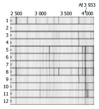
Methods: Surface enhanced laser desorption/ionization time-of-flight (SELDI-TOF) mass spectrometry was used to discover biomarkers for differentiating HBV induced LC from non-cirrhotic cohorts. A training population of 25 patients with HBV-induced LC, 20 patients with HCC, and 25 closely age-matched healthy men, was studied.
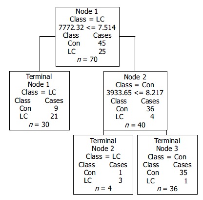
Results: Two biomarkers with M(r) 7 772 and 3 933 were detected in sera of non-cirrhotic cohorts, but not in patients with HBV-induced LC. A sensitivity of 80% for all LC patients, a specificity of 81.8% for all non-cirrhotic cohorts and a positive predictive value of 75% for the study population were obtained.
Conclusion: These two serum biomarkers for HBV-induced LC might be used for diagnosis and assessment of disease progression.
Figures

Similar articles
-
Surface enhanced laser desorption/ionization profiling: New diagnostic method of HBV-related hepatocellular carcinoma.J Gastroenterol Hepatol. 2009 Jan;24(1):55-62. doi: 10.1111/j.1440-1746.2008.05580.x. Epub 2008 Sep 24. J Gastroenterol Hepatol. 2009. PMID: 18823443
-
Prediction of chronic hepatitis B, liver cirrhosis and hepatocellular carcinoma by SELDI-based serum decision tree classification.J Cancer Res Clin Oncol. 2007 Nov;133(11):825-34. doi: 10.1007/s00432-007-0224-y. Epub 2007 May 22. J Cancer Res Clin Oncol. 2007. PMID: 17516088 Free PMC article.
-
Potential Serum Markers for Monitoring the Progression of Hepatitis B Virus-Associated Chronic Hepatic Lesions to Liver Cirrhosis.Gut Liver. 2015 Sep 23;9(5):665-71. doi: 10.5009/gnl14212. Gut Liver. 2015. PMID: 25963079 Free PMC article.
-
Natural history of chronic hepatitis B REVEALed.J Gastroenterol Hepatol. 2011 Apr;26(4):628-38. doi: 10.1111/j.1440-1746.2011.06695.x. J Gastroenterol Hepatol. 2011. PMID: 21323729 Review.
-
Novel biomarkers for HBV including point-of-care.Clin Liver Dis (Hoboken). 2024 Jun 28;23(1):e0205. doi: 10.1097/CLD.0000000000000205. eCollection 2024 Jan-Jun. Clin Liver Dis (Hoboken). 2024. PMID: 38952698 Free PMC article. Review. No abstract available.
Cited by
-
Screening serum hepatocellular carcinoma-associated proteins by SELDI-based protein spectrum analysis.World J Gastroenterol. 2008 Feb 28;14(8):1257-62. doi: 10.3748/wjg.14.1257. World J Gastroenterol. 2008. PMID: 18300354 Free PMC article.
-
Two classifiers based on serum peptide pattern for prediction of HBV-induced liver cirrhosis using MALDI-TOF MS.Biomed Res Int. 2013;2013:814876. doi: 10.1155/2013/814876. Epub 2013 Feb 19. Biomed Res Int. 2013. PMID: 23509784 Free PMC article.
-
Matrix stiffness-mediated effects on stemness characteristics occurring in HCC cells.Oncotarget. 2016 May 31;7(22):32221-31. doi: 10.18632/oncotarget.8515. Oncotarget. 2016. PMID: 27050147 Free PMC article.
-
Human body fluid proteome analysis.Proteomics. 2006 Dec;6(23):6326-53. doi: 10.1002/pmic.200600284. Proteomics. 2006. PMID: 17083142 Free PMC article. Review.
-
The current state of proteomics in GI oncology.Dig Dis Sci. 2009 Mar;54(3):431-57. doi: 10.1007/s10620-008-0656-5. Epub 2008 Dec 23. Dig Dis Sci. 2009. PMID: 19104933 Free PMC article. Review.
References
-
- Lai CL, Lok A, Wu PC, Ng M. Risk factors and hepatocellular cancer. Lancet. 1985;2:329–330. - PubMed
-
- Colombo M, de Franchis R, Del Ninno E, Sangiovanni A, De Fazio C, Tommasini M, Donato MF, Piva A, Di Carlo V, Dioguardi N. Hepatocellular carcinoma in Italian patients with cirrhosis. N Engl J Med. 1991;325:675–680. - PubMed
-
- Poniachik J, Bernstein DE, Reddy KR, Jeffers LJ, Coelho-Little ME, Civantos F, Schiff ER. The role of laparoscopy in the diagnosis of cirrhosis. Gastrointest Endosc. 1996;43:568–571. - PubMed
-
- Issaq HJ, Veenstra TD, Conrads TP, Felschow D. The SELDI-TOF MS approach to proteomics: protein profiling and biomarker identification. Biochem Biophys Res Commun. 2002;292:587–592. - PubMed
-
- He QY, Lau GK, Zhou Y, Yuen ST, Lin MC, Kung HF, Chiu JF. Serum biomarkers of hepatitis B virus infected liver inflammation: a proteomic study. Proteomics. 2003;3:666–674. - PubMed
Publication types
MeSH terms
Substances
LinkOut - more resources
Full Text Sources
Medical
