

E-cadherin and calretinin as immunocytochemical markers to differentiate malignant from benign serous effusions
- PMID: 15285029
- PMCID: PMC4576298
- DOI: 10.3748/wjg.v10.i16.2406
E-cadherin and calretinin as immunocytochemical markers to differentiate malignant from benign serous effusions
Abstract
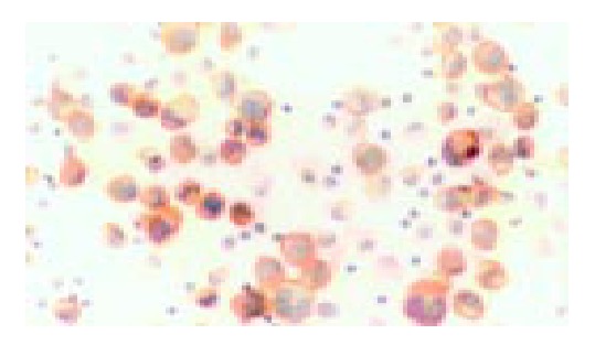
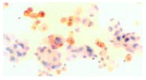
Aim: To investigate the expressions of E-cadherin and calretinin in exfoliated cells of serous effusions and evaluate their values in distinguishing malignant effusions from benign ones.
Methods: Fresh serous effusion specimens were centrifuged and exfoliated cells were collected. Cells were then processed with a standardized procedure, including paraformaldehyde fixation, BSA-PBS solution washing and smears preparation. E-cadherin and calretinin were detected by immunocytochemistry (ICC).
Results: In the exfoliated cells of serous effusions, most of carcinoma cells only expressed E-cadherin, and most of mesothelial cells only expressed calretinin, and benign cells (lymphocytes and granulocytes) did not express either of them. For E-cadherin, 85.7% (30/35) of malignant effusions and 8.1% (3/37) of benign fluids were ICC-positive (P<0.001). The sensitivity of E-cadherin ICC in the diagnosis of malignant effusions was 85.7%, specificity 91.9%, and diagnostic rate 88.9%. For calretinin, 94.6% (35/37) of benign effusions and 11.4% (4/35) of malignant effusions were ICC-positive (P<0.001). The sensitivity of calretinin ICC in the diagnosis of benign effusions was 94.6%, specificity 88.6%, and diagnostic rate 91.7%. For diagnosis of benign and malignant effusions by combining E-cadherin ICC and calretinin ICC, the specificities were up to 100% and 97.1%, respectively.
Conclusion: E-cadherin ICC and calretinin ICC are sensitive and specific in differential diagnosis of benign and malignant serous effusion specimens and specificities are evidently improved when both markers are combined.
Figures
References
-
- Chhieng DC, Yee H, Schaefer D, Cangiarella JF, Jagirdar J, Chiriboga LA, Jagirdar J, Chiriboga LA, Cohen JM. Calretinin staining pattern aids in the differentiation of mesothelioma from adenocarcinoma in serous effusions. Cancer. 2000;90:194–200. - PubMed
-
- Ascoli V, Scalzo CC, Taccogna S, Nardi F. The diagnostic value of thrombomodulin immunolocalization in serous effusions. Arch Pathol Lab Med. 1995;119:1136–1140. - PubMed
-
- Ascoli V, Carnovale-Scalzo C, Taccogna S, Nardi F. Utility of HBME-1 immunostaining in serous effusions. Cytopathology. 1997;8:328–335. - PubMed
-
- Morgan RL, De Young BR, McGaughy VR, Niemann TH. MOC-31 aids in the differentiation between adenocarcinoma and reactive mesothelial cells. Cancer. 1999;87:390–394. - PubMed
-
- Zimmerman RL, Fogt F, Goonewardene S. Diagnostic utility of BCA-225 in detecting adenocarcinoma in serous effusions. Anal Quant Cytol Histol. 2000;22:353–357. - PubMed
MeSH terms
Substances
LinkOut - more resources
Full Text Sources
