

Role of endoscopic miniprobe ultrasonography in diagnosis of submucosal tumor of large intestine
- PMID: 15285040
- PMCID: PMC4576308
- DOI: 10.3748/wjg.v10.i16.2444
Role of endoscopic miniprobe ultrasonography in diagnosis of submucosal tumor of large intestine
Abstract
Aim: To evaluate the role of miniprobe ultrasonography under colonoscope in the diagnosis of submucosal tumor of the large intestine, and to determine its imaging characteristics.
Methods: Thirty-five patients with submucosal tumors of the large intestine underwent miniprobe ultrasonography under colonoscope. The diagnostic results of miniprobe ultrasonography were compared with pathological findings of specimens by biopsy and surgical resection.
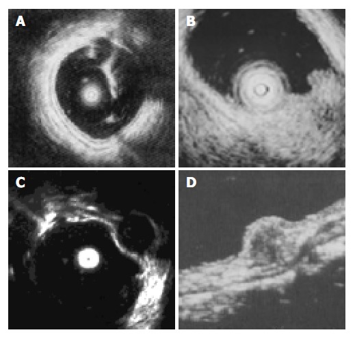
Results: Lipomas were visualized as hyperechoic homogeneous masses located in the submucosa with a distinct border. Leiomyomas were visualized as hypoechoic homogeneous mass originated from the muscularis propria. Leiomyosarcomas were shown with inhomogeneous echo and irregular border. Carcinoids were presented as submucosal hypoechoic masses with homogenous echo and distinct border. Lymphangiomas were shown as submocosal hypoechoic masses with cystic septal structures. Malignant lymphomas displayed as hypoechoic masses from mucosa to muscularis propria, while pneumatosis cystoids intestinalis originated from submucosa with a special sonic shadow. One large leiomyoma was misdiagnosed as leiomyosarcoma.
Conclusion: Endoscopic miniprobe ultrasonography can provide precise information about the size, layer of origin, border of submucosal tumor of the large intestine and has a high accuracy in the diagnosis of submucosal tumor of the large intestine. Pre-operative miniprobe ultrasonography under colonoscope may play an important role in the choice of therapy for submucosal tumor of the large intestine.
Figures
References
-
- Stergiou N, Haji-Kermani N, Schneider C, Menke D, Köckerling F, Wehrmann T. Staging of colonic neoplasms by colonoscopic miniprobe ultrasonography. Int J Colorectal Dis. 2003;18:445–449. - PubMed
-
- Waxman I, Saitoh Y, Raju GS, Watari J, Yokota K, Reeves AL, Kohgo Y. High-frequency probe EUS-assisted endoscopic mucosal resection: a therapeutic strategy for submucosal tumors of the GI tract. Gastrointest Endosc. 2002;55:44–49. - PubMed
-
- Takada N, Higashino M, Osugi H, Tokuhara T, Kinoshita H. Utility of endoscopic ultrasonography in assessing the indications for endoscopic surgery of submucosal esophageal tumors. Surg Endosc. 1999;13:228–230. - PubMed
-
- Roseau G, Dumontier I, Palazzo L, Chapron C, Dousset B, Chaussade S, Dubuisson JB, Couturier D. Rectosigmoid endometriosis: endoscopic ultrasound features and clinical implications. Endoscopy. 2000;32:525–530. - PubMed
MeSH terms
LinkOut - more resources
Full Text Sources
