. 2004 Nov 15;384(Pt 1):179-90.
doi: 10.1042/BJ20040658.

Guanidinium chloride denaturation of the dimeric Bacillus licheniformis BlaI repressor highlights an independent domain unfolding pathway

Affiliations

Guanidinium chloride denaturation of the dimeric Bacillus licheniformis BlaI repressor highlights an independent domain unfolding pathway

Christelle Vreuls et al. Biochem J. .

Abstract

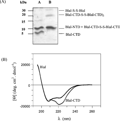
The Bacillus licheniformis 749/I BlaI repressor is a prokaryotic regulator that, in the absence of a beta-lactam antibiotic, prevents the transcription of the blaP gene, which encodes the BlaP beta-lactamase. The BlaI repressor is composed of two structural domains. The 82-residue NTD (N-terminal domain) is a DNA-binding domain, and the CTD (C-terminal domain) containing the next 46 residues is a dimerization domain. Recent studies have shown the existence of the monomeric, dimeric and tetrameric forms of BlaI in solution. In the present study, we analyse the equilibrium unfolding of BlaI in the presence of GdmCl (guanidinium chloride) using different techniques: intrinsic and ANS (8-anilinonaphthalene-l-sulphonic acid) fluorescence, far- and near-UV CD spectroscopy, cross-linking, analytical ultracentrifugation, size exclusion chromatography and NMR spectroscopy. In addition, the intact NTD and CTD were purified after proteolysis of BlaI by papain, and their unfolding by GdmCl was also studied. GdmCl-induced equilibrium unfolding was shown to be fully reversible for BlaI and for the two isolated fragments. The results demonstrate that the NTD and CTD of BlaI fold/unfold independently in a four-step process, with no significant co-operative interactions between them. During the first step, the unfolding of the BlaI CTD occurs, followed in the second step by the formation of an 'ANS-bound' intermediate state. Cross-linking and analytical ultracentrifugation experiments suggest that the dissociation of the dimer into two partially unfolded monomers takes place in the third step. Finally, the unfolding of the BlaI NTD occurs at a GdmCl concentration of approx. 4 M. In summary, it is shown that the BlaI CTD is structured, more flexible and less stable than the NTD upon GdmCl denaturation. These results contribute to the characterization of the BlaI dimerization domain (i.e. CTD) involved in the induction process.


Figures

Figure 1
Figure 1. β-Lactamase induction mechanism in B. licheniformis 749/I
The B. licheniformis BlaP β-lactamase is inducible by a β-lactam antibiotic (the inducer) [2]. The regulation of β-lactamase production involves three regulatory genes, blaI, blaR1 and blaR2. The first two genes encode a repressor and a penicillin-sensory transducer respectively ([4], but see [4a]), whereas the third gene is not identified yet. (A) In the absence of a β-lactam antibiotic, the BlaI repressor is bound as a dimer to three operator sequences (OP1, OP2 and OP3) located in the intergenic region between blaP and blaI-blaR operon. Bound to DNA, BlaI prevents the transcription of the blaP, blaI and blaR1 genes ([4], but see [4a]), [5]. (B) When a β-lactam antibiotic is added in the medium, the extracellular penicillin-binding domain of BlaR (BlaR-CTD) is acylated by the antibiotic [6], and a signal is transduced through the transmembrane segment resulting in the activation of the intracellular metalloprotease domain by proteolysis [7]. In B. licheniformis, it has been postulated that the activated metalloprotease converts a pro-coactivator into a co-activator, whose final target is BlaI itself. The BlaI dimer complexed with the co-activator is then released from its operator and β-lactamase is produced at high level ([4], but see [4a]). The product of blaR2 is necessary for the induction process; it is postulated that it can be involved in the activation of the intracellular domain of the BlaR receptor or in the production of the pro-coactivator [3].
Figure 2
Figure 2. Views of B. licheniformis BlaI-NTD and S. aureus MecI
(A) B. licheniformis BlaI-NTD (residues 1–82) belongs to the winged-helix subfamily and consists of a three-stranded β-sheet (S1, Ser23–Asn25; S2, Leu57–Glu62; S3, Val65–Pro70) packed against three α-helices (H1, Asp9–Lys20; H2, Thr26–Thr36; H3, Pro41–Lys53) arranged in the order H1-S1-H2-T1-H3-S2-W1-S3 [11]. S2 and S3 form an antiparallel hairpin (loop called wing W1: Gly63–Arg64) and S1 is connected in parallel with S3. T1 (Ser37–Ser40) is a type 1 turn connecting H2 and H3. The α-helices are in yellow the β-sheet is in red and the tryptophan residues are in black. (B) The S. aureus MecI dimer is composed of two independent winged-helix domains and of two intertwining dimerization domains. The overall fold topology of the NTD is H1-H2-H3-S1-W1-S2, and the CTD is composed of three consecutive α-helices H4-H5-H6 held together by two hydrophobic cores between H4 and H6 [13]. The first monomer is in blue. The NTD of the second monomer (residues 1–73) is in the same colours as BlaI-NTD, and the CTD (residues 74–123) is in green. The S. aureus BlaI and MecI repressors are 60% identical when compared with each other, and 31–41% identical when compared with the B. licheniformis BlaI repressor [14].
Figure 3
Figure 3. Characterization of the BlaI CTD
(A) Study of the oligomerization state of the BlaI CTD by cross-linking experiments. Purified BlaI was subjected to papain digestion. Cross-linking was performed by mixing 20 μmol of the digested protein with 50 μmol of DSP, and the results were analysed by SDS/PAGE (see the Materials and methods section). Lane A corresponds to the digested protein without DSP, and lane B to the digested protein incubated with DSP. Lane B highlights the dimeric (10 kDa) and tetrameric (20 kDa) forms of the BlaI CTD. (B) Far-UV CD spectra of BlaI and BlaI-CTD(His)6. Proteins were incubated in 50 mM phosphate buffer, pH 7.6, supplemented with 200 mM KCl and 13 mM CHAPS for BlaI-CTD(His)6. The protein concentrations used were 0.5 mg·ml−1 in a 0.1 cm cell.
Figure 4
Figure 4. Study of the reversibility of the BlaI repressor GdmCl-induced denaturation by band-shift assays
(A) Binding of BlaI to the OP1 target under native conditions (0 M GdmCl). ‘Free’ and ‘Cp’ represent the free DNA target and the DNA–protein complex respectively. The fluorescence is expressed in arbitrary units. DNA probe OP1 (0.5×10⁻⁸ M) was incubated with purified BlaI repressor (3.3×10⁻⁸ M) and treated as described previously [16]. (B) Binding of BlaI to the OP1 target after denaturation–renaturation of the protein. Protein and DNA concentrations are the same as in (A).
Figure 5
Figure 5. BlaI and BlaI-NTD intrinsic fluorescence emission spectra at various GdmCl concentrations
Spectra were recorded at 25 °C. Proteins (2 μM) were in 50 mM phosphate, pH 7.6 buffer supplemented with 200 mM KCl. The excitation wavelength was 280 nm. (A) Fluorescence emission spectra of BlaI under native and unfolded conditions (0 M and 5 M GdmCl (‘GdnHCl’) respectively). A.U., arbitrary units. (B) Variation of the λmax of BlaI (○) and BlaI-NTD (•) as a function of GdmCl concentration.
Figure 6
Figure 6. ANS-bound fluorescence emission spectra of BlaI, BlaI-NTD and BlaI-CTD(His)6
Changes of the emission λmax of ANS bound to (A) BlaI, (B) BlaI-NTD and (C) BlaI-CTD(His)6 as a function of GdmCl (‘GdnHCl’) concentration. Spectra were recorded at 25 °C. Proteins (25 μM) were incubated in 50 mM phosphate buffer, pH 7.6, supplemented with 200 mM KCl and also 13 mM CHAPS for BlaI-CTD(His)6. The ANS concentration was 500 μM. The excitation wavelength was 350 nm.
Figure 7
Figure 7. CD studies of BlaI and BlaI-NTD
Far-UV CD spectra of (A) BlaI and (B) BlaI-NTD under native conditions (50 mM phosphate buffer, pH 7.6, supplemented with 200 mM KCl) and in 5 M GdmCl (‘GdnHCl’) (same buffer). The protein concentration was 0.5 mg·ml−1 in a 0.1 cm cell. The inset shows GdmCl-induced unfolding transition followed at 222 nm. (C) Near-UV CD spectra of BlaI under native conditions and in 5 M GdmCl (same buffers). The protein concentration was 1 mg·ml−1 in a 1 cm cell. The inset shows GdmCl-induced unfolding transition followed at 282 nm.
Figure 8
Figure 8. Two-dimensional 1H/15N-HSQC NMR spectra of BlaI and BlaI-NTD
(A) BlaI (0.4 mM) and (B) BlaI-NTD (0.3 mM) were in 50 mM phosphate buffer, pH 7.6, supplemented with 200 mM KCl and the appropriate GdmCl concentration: 0 M GdmCl (upper left panels), 1 M GdmCl (upper right panels) and 5 M GdmCl (lower right panels). The latter spectra (lower left panels) demonstrate the full renaturation of the unfolded proteins after removal of the denaturant (for further details, see the text and the Materials and methods section). A decrease of peak intensity was observed for all 1H/15N cross-peaks upon increasing the GdmCl concentration. This was due to the change in the quality factor of the probe (Q factor) linked to the presence of high amounts of salt [29].
Figure 9
Figure 9. Effect of GdmCl (‘GdnHCl’) on the oligomerization state of BlaI
(A) SDS/PAGE after covalent cross-linking of the BlaI protein at increasing GdmCl concentrations. Protein (20 μM) was incubated for 2 h with DSP and subsequently the reaction was quenched 15 min on ice with 10 μl of 1 M Tris, as described in the Materials and methods section. Samples were electrophoresed on an SDS/4–15% acrylamide gel. Shown are standard markers (lane 1), BlaI after cross-linking in the absence of GdmCl (lane 2) and protein pre-incubated in a GdmCl concentration of 1, 2, 3 or 4 M before cross-linking (lanes 3–6 respectively). (B) Sedimentation velocity experiments: GdmCl concentration-dependence of the sedimentation coefficients s20,w (S). BlaI (60 μM) was incubated in 50 mM Hepes buffer, pH 7.6 containing 200 mM KCl, 1 mM EDTA, 5% glycerol and supplemented with the required GdmCl concentration. The samples were centrifuged at 40000 rev./min and scans were collected every 15 min at a wavelength of 280 nm. Sedimentation coefficients were calculated according to the method of Stafford (see [20]).
Figure 10
Figure 10. SEC elution profiles of BlaI
BlaI (50 μM) was incubated for 2 h in 50 mM Hepes buffer, pH 7.6, containing 200 mM KCl, 1 mM EDTA, 5% glycerol and supplemented with the required GdmCl (‘GdnHCl’) concentration, and then deposited on to a Superdex 75 column pre-equilibrated with the same GdmCl concentration. Other conditions were as described in the Materials and methods section. (A) Elution profiles of BlaI at different GdmCl concentrations. (B) Rs of BlaI as a function of GdmCl concentration. D, I, M and U represent respectively the native dimeric protein, the ‘ANS-bound’ intermediate state, the partially unfolded monomer and the unfolded BlaI protein.
Scheme 1
Scheme 1. Representation of the GdmCl-induced unfolding pathway of the BlaI repressor
BlaI denaturation proceeds through the independent sequential unfolding of the two domains and involves three distinct intermediate states: (i) an altered dimer (D') with its CTD in a random state and its NTD in a native conformation; (ii) a partially unfolded ‘ANS-bound dimeric intermediate state’ with highly exposed hydrophobic surfaces; and (iii) a partially unfolded monomer (M) with an intact NTD. It should be noted that the NTD remains folded in all the intermediate states, showing the high stability of this independent domain.

Similar articles

Cited by

References

    1. Charlier P., Coyette J., Dehareng D., Dive G., Duez C., Dusart J., Fonze E., Fraipont C., Frère J. M., Galleni M., et al. Résistance bacterienne aux β-lactamases. Medecine/sciences. 1998;14:544–549.
    1. Joris B., Hardt K., Ghuysen J. M. Induction of β-lactamase and low-affinity penicillin binding protein 2′ synthesis in Gram-positive bacteria. New Compr. Biochem. 1994;27:505–515.
    1. Filée P., Benlafaya K., Delmarcelle M., Moutzourelis G., Frère J. M., Brans A., Joris B. The fate of the BlaI repressor during the induction of the Bacillus licheniformis BlaP β-lactamase. Mol. Microbiol. 2002;44:685–694. - PubMed
    1. Himeno T., Imanaka T., Aiba S. Nucleotide sequence of the penicillinase repressor gene penI of Bacillus licheniformis and regulation of penP and penI by the repressor. J. Bacteriol. 1986;168:1128–1132. - PMC - PubMed
    1. Erratum. J. Bacteriol. 1987;169:3392.

Publication types

MeSH terms