Review
. 2004 Aug;4(8):583-94.
doi: 10.1038/nri1412.

Fibrotic disease and the T(H)1/T(H)2 paradigm

Affiliations
Review

Fibrotic disease and the T(H)1/T(H)2 paradigm

Thomas A Wynn. Nat Rev Immunol. 2004 Aug.

Abstract

Tissue fibrosis (scarring) is a leading cause of morbidity and mortality. Current treatments for fibrotic disorders, such as idiopathic pulmonary fibrosis, hepatic fibrosis and systemic sclerosis, target the inflammatory cascade, but they have been widely unsuccessful, largely because the mechanisms that are involved in fibrogenesis are now known to be distinct from those involved in inflammation. Several experimental models have recently been developed to dissect the molecular mechanisms of wound healing and fibrosis. It is hoped that by better understanding the immunological mechanisms that initiate, sustain and suppress the fibrotic process, we will achieve the elusive goal of targeted and effective therapeutics for fibroproliferative diseases.


Figures
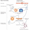
Figure 1
Figure 1. The pathogenesis of fibrotic disease
Healing is the normal reaction of tissues after injury. Damaged epithelial and/or endothelial cells release inflammatory mediators that initiate an antifibrinolytic–coagulation cascade, which triggers blood-clot formation. Next, epithelial and endothelial cells secrete growth factors and chemokines that stimulate the proliferation and recruitment of leukocytes that produce pro-fibrotic cytokines, such as interleukin-13 (IL-13) and transforming growth factor-β (TGF-β). Stimulated myofibroblasts and epithelial/endothelial cells also produce matrix metalloproteinases (MMPs), which disrupt the basement membrane, allowing the efficient recruitment of cells to sites of injury. After this migration, activated macrophages and neutrophils ‘clean-up’ tissue debris and dead cells. They also produce cytokines and chemokines that recruit and activate T cells, which are important components of granulation tissue as they secrete pro-fibrotic cytokines (such as IL-13). Fibroblasts are subsequently recruited and activated. Fibroblasts can be derived from local mesenchymal cells or recruited from the bone marrow (known as fibrocytes). Epithelial cells can undergo epithelial–mesenchymal transition, providing a rich renewable source of fibroblasts. Revascularization of the wound also occurs at this time. After activation, myofibroblasts cause wound contraction, the process in which the edges of the wound migrate towards the centre. Last, epithelial and/or endothelial cells divide and migrate over the basal layers to regenerate the epithelium or endothelium, respectively, which completes the healing process. However, when repeated injury occurs, chronic inflammation and repair can cause an excessive accumulation of extracellular-matrix components, such as the collagen that is produced by fibroblasts, and lead to the formation of a permanent fibrotic scar. Pro-fibrotic mediators, such as IL-13 and TGF-β, amplify these processes. The net amount of collagen deposited by fibroblasts is regulated by continued collagen synthesis and collagen catabolism. The degradation of collagen is controlled by MMPs and their inhibitors (such as tissue inhibitors of matrix metalloproteinases, TIMPs), and the net increase in collagen within a wound is controlled by the balance of these opposing mechanisms.
Figure 2
Figure 2. Opposing roles for TH1 and TH2 cytokines in fibrosis
The T helper 1 (TH1)-cell cytokine interferon-γ (IFN-γ) directly suppresses collagen synthesis by fibroblasts. It achieves this through regulating the balance of matrix metalloproteinase (MMP) and tissue inhibitor of matrix metalloproteinase (TIMP) expression, thereby controlling the rates of collagen degradation and synthesis, respectively, in the extracellular matrix. IFN-γ and/or interleukin-12 (IL-12) might also indirectly inhibit fibrosis by reducing pro-fibrotic cytokine expression by TH2 cells. The main TH2 cytokines (IL-4, IL-5 and IL-13) enhance collagen deposition by various mechanisms; however, IL-13 seems to be the crucial mediator.
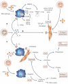
Figure 3
Figure 3. IL-13 and TGF-β might function independently or cooperatively to promote collagen deposition by fibroblasts
Interleukin-13 (IL-13) promotes collagen production by three distinct but possibly overlapping mechanisms. a | IL-13 is produced by activated CD4+ T helper 2 (TH2) cells and stimulates the production of latent transforming growth factor-β (TGF-β) by macrophages. The activation of latent TGF-β is important for both its physiological and pathological actions on target cells, such as fibroblasts. Following the intracellular processing of prepro-TGF-β to its latent complex, the latent TGF-β is secreted and then anchored to the macrophage membrane through thrombospondin-1 bound to its receptor, CD36. Activation of TGF-β might be mediated by plasmin/serine protease- and/or matrix metalloproteinase 9 (MMP9)-dependent mechanisms. After the latency-associated peptide (LAP) is cleaved, TGF-β is free to bind and activate TGF-β receptors (TGF-βRs) expressed by fibroblasts. In this pathway, the fibrogenic effects of IL-13 are mediated largely by the downstream actions of TGF-β. b | Because fibroblasts express IL-13 receptors (IL-13Rs), IL-13 might also directly activate the collagen-producing machinery in fibroblasts-. c | IL-13 can also promote the alternative activation of macrophages and/or fibroblasts. By upregulating arginase activity in these cells, IL-13 increases l-ornithine, l-proline and polyamine concentrations, which promotes fibroblast proliferation, collagen production and ultimately, fibrosis. Interferon-γ (IFN-γ) produced by TH1 cells seems to antagonize all of these pathways. IFN-γ inhibits alternative macrophage activation, inducing nitric-oxide synthase 2 (NOS2) instead of arginase, and directly decreases collagen synthesis by fibroblasts. NOS2 activity promotes the production of l-hydroxyarginine, l-citrulline and nitric oxide. The intermediate by product l-hydroxyarginine also functions as a potent inhibitor of arginase activity, which can further antagonize the fibrotic pathway. OAT, ornithine amino transferase; ODC, ornithine decarboxylase.
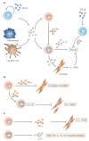
Figure 4
Figure 4. Regulatory T cells, IL-10 and IL-13Rα2 function as endogenous inhibitors of tissue fibrosis
a | Recent studies indicate that CD4+CD25+ regulatory T (TReg) cells and non-T cells that express interleukin-10 (IL-10) (possibly macrophages and/or dendritic cells) cooperate to generate polarized T helper 2 (TH2)-cell responses. Transforming growth factor-β1 (TGF-β1)-producing TReg cells might also promote the development of IL-10-producing TReg cells. IL-10 can directly inhibit collagen synthesis by fibroblasts. IL-10 also inhibits interferon-γ (IFN-γ) production by TH1 cells, while promoting the development of a polarized but controlled TH2 response. In this setting, IL-13 induces collagen deposition by fibroblasts; however, it also induces expression of its decoy receptor IL-13 receptor-α2 (IL-13Rα2), which ultimately attenuates the response. Recent evidence indicates that fibroblasts are an important source of IL-13Rα2 (refs 109-112). Therefore, both IL-10 produced by TReg cells and the IL-13Rα2 might cooperate to control fibrosis during polarized TH2 responses. b | When a highly polarized TH1 response is generated, little IL-13 is produced. Consequently, fibrosis is minimal and decoy-receptor expression remains low. c | When a mixed TH1/TH2 response develops, IFN-γ might decrease production of the IL-13 decoy receptor and upregulate IL-13 effector function,,. In this case, although IL-13 concentrations might slightly decrease or remain unchanged, more IL-13 is free to bind the signalling receptor. This could explain the unusual tendency of mixed responses to trigger severe tissue pathology-.

References

    1. Cotran RS, Kumar V, Collins T. In: Pathologic Basis of Disease. Cotran RS, Kumar V, Collins T, editors. Vol. 6. W. B. Saunders Company; Philadelphia: 1999. pp. 89–112.
    1. Wynn TA, et al. An IL-12-based vaccination method for preventing fibrosis induced by schistosome infection. Nature. 1995;376:594–596. An early example of the opposing activities of TH1 and TH2 responses in a progressive fibrotic disease. - PubMed
    1. Hoffmann KF, Cheever AW, Wynn TA. IL-10 and the dangers of immune polarization: excessive type 1 and type 2 cytokine responses induce distinct forms of lethal immunopathology in murine schistosomiasis. J. Immunol. 2000;164:6406–6416. - PubMed
    1. Gurujeyalakshmi G, Giri SN. Molecular mechanisms of antifibrotic effect of interferon-γ in bleomycin-mouse model of lung fibrosis: downregulation of TGF-β and procollagen I and III gene expression. Exp. Lung Res. 1995;21:791–808. - PubMed
    1. Keane MP, Belperio JA, Burdick MD, Strieter RM. IL-12 attenuates bleomycin-induced pulmonary fibrosis. Am. J. Physiol. Lung Cell. Mol. Physiol. 2001;281:L92–L97. - PubMed