Comparative Study
. 2004 Aug;114(3):322-9.
doi: 10.1172/JCI21153.

Obstruction of extrahepatic bile ducts by lymphocytes is regulated by IFN-gamma in experimental biliary atresia

Affiliations
Comparative Study

Obstruction of extrahepatic bile ducts by lymphocytes is regulated by IFN-gamma in experimental biliary atresia

Pranavkumar Shivakumar et al. J Clin Invest. 2004 Aug.

Abstract

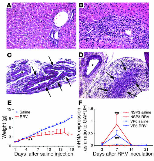
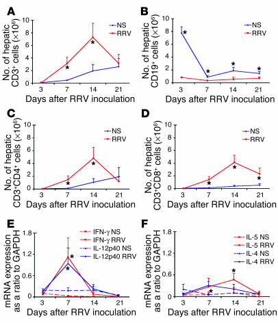
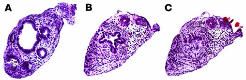
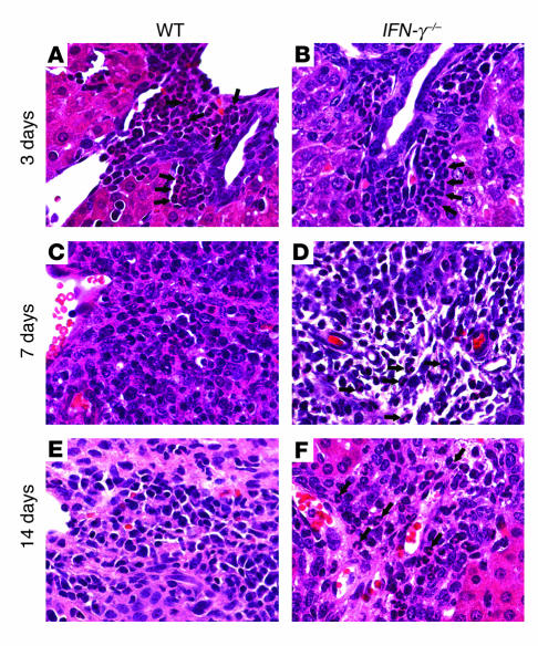
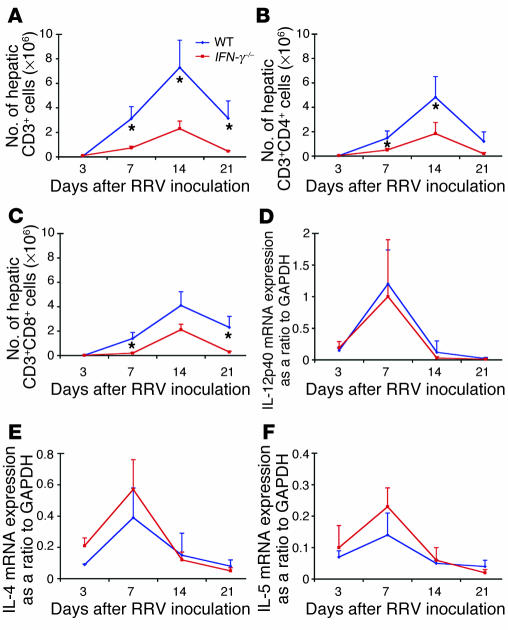
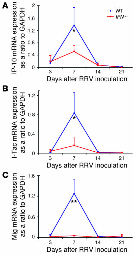
The etiology and pathogenesis of bile duct obstruction in children with biliary atresia are largely unknown. We have previously reported that, despite phenotypic heterogeneity, genomic signatures of livers from patients display a proinflammatory phenotype. Here, we address the hypothesis that production of IFN-gamma is a key pathogenic mechanism of disease using a mouse model of rotavirus-induced biliary atresia. We found that rotavirus infection of neonatal mice has a unique tropism to bile duct cells, and it triggers a hepatobiliary inflammation by IFN-gamma-producing CD4(+) and CD8(+) lymphocytes. The inflammation is tissue specific, resulting in progressive jaundice, growth failure, and greater than 90% mortality due to obstruction of extrahepatic bile ducts. In this model, the genetic loss of IFN-gamma did not alter the onset of jaundice, but it remarkably suppressed the tissue-specific targeting of T lymphocytes and completely prevented the inflammatory and fibrosing obstruction of extrahepatic bile ducts. As a consequence, jaundice resolved, and long-term survival improved to greater than 80%. Notably, administration of recombinant IFN-gamma led to recurrence of bile duct obstruction following rotavirus infection of IFN-gamma-deficient mice. Thus, IFN-gamma-driven obstruction of bile ducts is a key pathogenic mechanism of disease and may constitute a therapeutic target to block disease progression in patients with biliary atresia.


Figures

Figure 1
Figure 1
RRV infection induces biliary inflammation and growth failure in neonatal mice. WT Balb/c mice were injected with normal saline (control) or RRV within 24 hours of birth, and the hepatobiliary system was examined 7 days later. (A) While livers of control mice had normal appearance of the portal tracts, RRV challenge resulted in the expansion of portal spaces by inflammatory cells and proliferating bile duct cells (B). (C) Cross section of the extrahepatic bile duct of a control mouse revealed normal epithelium and unobstructed lumen (arrows). (D) In contrast, injection of RRV produced lumenal obstruction of extrahepatic bile ducts (arrows). Tissue sections were stained with H&E. Magnification of ×400 for A and B, ×200 for C and D. Single asterisks denote neighboring arteries in C and D. (E) It can be seen that RRV injection also led to poor growth during the suckling period. **P < 0.01 when compared with controls at days 7–16; n = 25 mice in the beginning of the experiment. Expression of mRNA encoding RRV nonstructural (NSP3) and structural (VP6) proteins was high at day 7 but (F) undetectable at day 14. **P < 0.01; n = 4–7 mice per group at each time point.
Figure 2
Figure 2
RRV targets the biliary epithelium in neonatal mice. Fluorescence immunostaining shows specific signal for RRV (red, A) and the cholangiocyte cytoskeletal filament CK7 (green, B) in the epithelium of extrahepatic bile ducts 3 days after RRV inoculation, with colocalization of RRV and CK7 (yellow, C).
Figure 3
Figure 3
RRV infection results in Th1 polarization of hepatic lymphocytes. Hepatic cell surface staining by flow cytometry for CD3 (A) and CD19 (B) shows that the lymphocytic infiltrate in portal tracts is predominantly composed of CD3+ cells beginning 7 days after RRV inoculation. CD3+ cells also showed staining with CD4+ (C) or CD8+ (D). (E) Functional polarization of T lymphocytes is demonstrated 7 days after RRV challenge by an increase in mRNA expression for IFN-γ and IL-12p40. (F) The mRNA expression for Th2 cytokines also increases above the levels of controls at 7 and 14 days, but at lower levels when compared with Th1 cytokines. *P < 0.05 when the RRV group is compared with controls; n = 4–7 mice per group at each time point. NS, normal saline.
Figure 4
Figure 4
Loss of IFN-γ improves symptoms of biliary obstruction after RRV challenge. Inoculation of RRV into newborn WT (A) mice induced jaundice, acholic stools, and bilirubinuria in all mice by 7 days, which persisted for the duration of the study in approximately 80% of mice. Although these symptoms also developed in IFN-γ–/– mice in a timely fashion (B), jaundice, acholic stools, and bilirubinuria resolved between 8 and 13 days after RRV challenge. n = 17 for WT mice; n = 12 for IFN-γ–/– mice.
Figure 5
Figure 5
Loss of IFN-γ prevents obstruction of extrahepatic bile ducts. Anatomical view of the hilum (AD) of WT Balb/c mice displayed small, edematous gallbladders (*) at 7 and 14 days after RRV challenge, with long- (7 days) or short- (14 days) segment atresia of extrahepatic bile ducts (thin arrows). In contrast, IFN-γ–/– mice displayed gallbladders distended with bile (**) and unobstructed bile ducts (thick arrows). Arrowheads point to arterial vessels that follow extrahepatic bile ducts. Microscopically (EH), bile ducts of WT Balb/c mice demonstrated lumenal obstruction by inflammatory cells (7 days) and extracellular matrix (14 days). In IFN-γ–/– mice, extrahepatic bile ducts had periductal inflammation and mild epithelial injury, but the lumen remained patent and without accumulation of matrix substrates at 7–14 days. Sections were stained with H&E; magnification, ×200; arrows in (E) and (G) denote obstructed bile ducts.
Figure 6
Figure 6
Administration of recombinant IFN-γ results in obstruction of extrahepatic bile ducts in IFN-γ–/– mice. H&E staining of transverse sections along the extrahepatic bile duct of an IFN-γ–/– mouse that received daily intraperitoneal injections of 2,000 U of recombinant IFN-γ per gram body weight following RRV challenge. (A) A patent proximal segment of the duct. (B) A narrow lumen with increasing periductal inflammation (approximately 700 μm from the section in A). (C) Section approximately 100 μm from the section in B; the inflammation completely occludes the extrahepatic bile duct. Magnification ×200.
Figure 7
Figure 7
Persistent infiltration of portal space by neutrophils in IFN-γ–/– mice. RRV challenge induces a neutrophil-based pericholangitis within 3 days in WT and IFN-γ–/– mice (A and B). In WT mice, inflammatory cells switch to lymphocytes in expanded portal spaces 7–14 days after challenge (C and E). This switch is incomplete in IFN-γ–/– mice, which continue to display portal neutrophils (D and F). Arrows point to neutrophils.
Figure 8
Figure 8
Hepatic population of T lymphocytes and cytokine expression in IFN-γ–/– mice. Flow cytometry shows a decreased population in the livers of IFN-γ–/– mice by CD3+CD4+ and CD3+CD8+ lymphocytes after RRV challenge (AC). Despite the loss of IFN-γ, expression of hepatic mRNA expression for IL-2p40, IL-4, and IL-5 in response to RRV challenge does not change (DF). n = 4–7 mice per group at each time point. *P < 0.04.
Figure 9
Figure 9
Impaired expression of chemokines following RRV challenge of IFN-γ–/– mice. Hepatic mRNA expression for downstream targets of IFN-γ as a ratio to GAPDH shows a lower increase for IP-10 (A) and I-Tac (B) 7 days after RRV challenge, and a complete suppression of Mig (C). *P < 0.05 and **P < 0.001 when WT is compared to IFN-γ–/– mice; n = 4–7 mice for each group and at all time points.

Similar articles

Cited by

References

    1. Balistreri WF, et al. Biliary atresia: current concepts and research directions. Summary of a symposium. Hepatology. 1996;23:1682–1692. - PubMed
    1. Perlmutter DH, Shepherd RW. Extrahepatic biliary atresia: a disease or a phenotype? Hepatology. 2002;35:1297–1304. - PubMed
    1. Sokol RJ, Mack C, Narkewicz MR, Karrer FM. Pathogenesis and outcome of biliary atresia: current concepts. J. Pediatr. Gastroenterol. Nutr. 2003;37:4–21. - PubMed
    1. Bezerra JA, et al. Genetic induction of proinflammatory immunity in children with biliary atresia. Lancet. 2002;360:1563–1659. - PubMed
    1. Riepenhoff-Talty M, et al. Group A rotaviruses produce extrahepatic biliary obstruction in orally inoculated newborn mice. Pediatr. Res. 1993;33:394–399. - PubMed

Publication types

MeSH terms