. 2004 Aug;114(3):408-18.
doi: 10.1172/JCI21100.

Integrin engagement regulates monocyte differentiation through the forkhead transcription factor Foxp1

Affiliations

Integrin engagement regulates monocyte differentiation through the forkhead transcription factor Foxp1

Can Shi et al. J Clin Invest. 2004 Aug.

Abstract

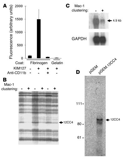
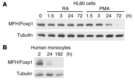
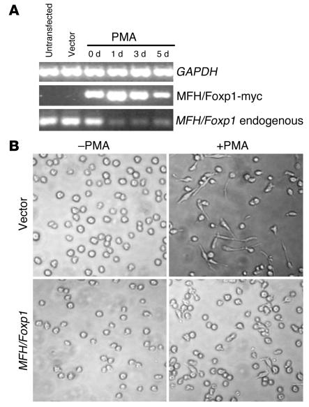
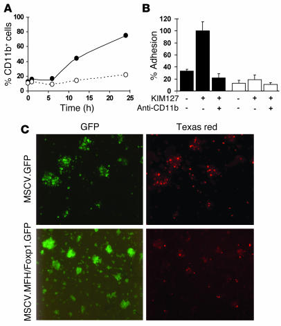
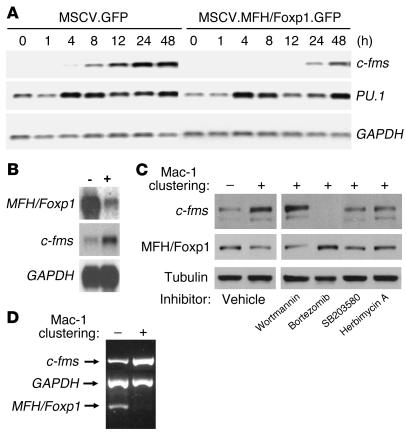
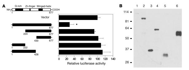
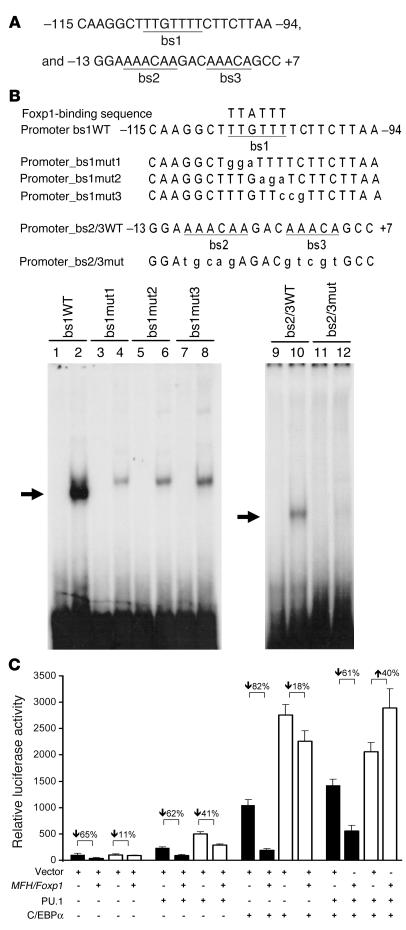
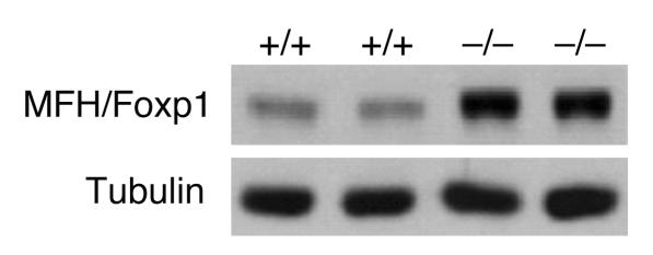
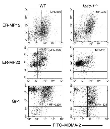
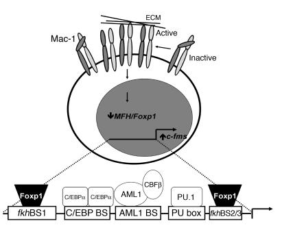
The precise signals responsible for differentiation of blood-borne monocytes into tissue macrophages are incompletely defined. "Outside-in" signaling by integrins has been implicated in modulation of gene expression that affects cellular differentiation. Herein, using differential display PCR, we have cloned an 85-kDa forkhead transcription factor (termed Mac-1-regulated forkhead [MFH] and found subsequently to be identical to Foxp1) that is downregulated in beta(2)-integrin Mac-1-clustered compared with Mac-1-nonclustered monocytic THP-1 cells. MFH/Foxp1 is expressed in untreated HL60 cells, and its expression was markedly reduced during phorbol ester-induced monocyte differentiation, but not retinoic acid-induced granulocyte differentiation. Overexpression of MFH/Foxp1 markedly attenuated phorbol ester-induced expression of c-fms, which encodes the M-CSF receptor and is obligatory for macrophage differentiation. This was accompanied by decreased CD11b expression, cell adhesiveness, and phagocytosis. Using electromobility shift and reporter assays, we have established that MFH/Foxp1 binds to previously uncharacterized sites within the c-fms promoter and functions as a transcriptional repressor. Deficiency of Mac-1 is associated with altered regulation of MFH/Foxp1 and monocyte maturation in vivo. Taken together, these observations suggest that Mac-1 engagement orchestrates monocyte-differentiation signals by regulating the expression of the forkhead transcription repressor MFH/Foxp1. This represents a new pathway for integrin-dependent modulation of gene expression and control of cellular differentiation.


Figures

Figure 1
Figure 1
Clustering of Mac-1 downregulates expression of 12CC4/MFH/Foxp1. (A) Clustering of Mac-1. Cytokine-treated THP-1 cells were added to fibrinogen-coated and gelatin-blocked wells. Adhesion was promoted by the addition of the anti-CD18 stimulating mAb KIM127 (5 μg/ml) in the presence and absence of the anti-CD11b mAb LPM19c (10 μg/ml). After washing, adhesion was quantified by measurement of the fluorescence of 2′,7′-bis-(2-carboyethyl)-5-(and-6)-carboxyfluorescein, acetoxymethyl ester–loaded (BCECF AM-loaded) THP-1 cells (Molecular Probes, Eugene, Oregon, USA). Triplicate determination (mean ± SD) representative of three separate experiments is shown. (B) Clustering of Mac-1 downregulates expression of 12CC4. Mac-1–dependent gene expression was examined using DD-PCR with RNA from Mac-1–clustered (+) and –nonclustered (–) THP-1 cells. cDNA expression patterns were analyzed from three independent clustering experiments. The arrow designates a band (12CC4) with reduced intensity after clustering in all three experiments. (C) Northern analysis. Northern blots containing 1 μg/lane of mRNA isolated from Mac-1–clustered and –nonclustered THP-1 cells were probed with 32P-labeled 12CC4 cDNA. RNA size is indicated in kilobases. (D) In vitro transcription and translation of vector alone and 12CC4. In vitro–coupled transcription and translation of native full-length 12CC4 (4,954 bp) produced a single band of predicted size (molecular weight ∼85,000).
Figure 2
Figure 2
Monocyte differentiation and expression of MFH/Foxp1. (A) HL60 cells. The expression of MFH/Foxp1 was examined in an in vitro system of myelomonocytic differentiation. HL60 cells were treated with PMA (10 nM) to induce monocytic differentiation (42) or with RA (100 nM) to induce granulocytic differentiation (22). (B) Human monocytes. MFH/Foxp1 expression was also assessed after adhesion-induced differentiation of human peripheral blood monocytes. At the indicated times, cells were lysed in situ with SDS-PAGE reduced-sample buffer and then immunoblotted sequentially with anti–MFH/Foxp1 and anti-tubulin antibodies.
Figure 3
Figure 3
Effect of overexpression of MFH on PMA-induced monocyte differentiation of HL60 cells. HL60 cells were stably transfected using pcDNA3.1/myc-his(-)A vector containing full-length MFH/Foxp1. (A) Semiquantitative PCR of endogenous MFH/Foxp1 and MFH/Foxp1-myc. (B) Light microscopy 12 hours after PMA treatment of HL60 cells.
Figure 4
Figure 4
Effect of overexpression of MFH on CD11b expression, cellular adhesion, and phagocytosis. (A) CD11b expression. HL60 cells, modified for MSCV retroviral transduction, were transduced with MSCV.MFH/Foxp1.GFP (open circles) or vector control (MSCV.GFP) (filled circles). Transduced HL60 cells were then treated with PMA (10 nM), and CD11b expression was assessed over time by FACS analysis using r-PE–conjugated anti-CD11b mAb (LPM19c). Data represent percentage CD11b-positive cells over time from three independent experiments. (B) Adhesion. Transduced HL60 cells (MSCV.GFP, black bars; MSCV.MFH/Foxp1.GFP, white bars) were harvested 24 hours after PMA treatment to investigate their adhesion to fibrinogen-coated microtiter wells. Adhesion was induced by the addition of KIM127. CD11b-dependence was examined by incubation of cells with the anti-CD11b mAb (LPM19c). Data represent percentage adhesion of vector control HL60 cells treated with the β2-integrin–stimulating mAb KIM127 (mean ± SD, n = 3). (C) Phagocytosis. GFP-expressing HL60 cells were cultured in the presence of Texas red–conjugated zymosan particles for 2 hours at 37°C as described in Methods. Cells were viewed for internalization of the particles by fluorescence microscopy after quenching of extracellular fluorescence with trypan blue. Assays were performed in triplicate from at least three independent experiments.
Figure 5
Figure 5
MFH/Foxp1and c-fms expression. (A) Overexpression of MFH/Foxp1attenuates expression of c-fms. HL60 cells transduced with control virus (MSCV.GFP) or MSCV.MFH/Foxp1.GFP were stimulated with PMA (10 nM), and the expression of c-fms and PU.1 over time was assessed by Northern analysis. (B) Clustering of Mac-1 enhances c-fms expression. Northern blots containing 1 μg/lane of mRNA isolated from Mac-1–clustered (+) and –nonclustered (–) THP-1 cells were probed with 32P-labeled c-fms and MFH/Foxp1 cDNA probes. (C) Regulation of MFH/Foxp1 and c-fms protein by Mac-1. MFH/Foxp1 and c-fms proteins were assessed after Mac-1 clustering. THP-1 cells were lysed 24 hours after clustering with SDS-PAGE reduced-sample buffer and then immunoblotted sequentially with anti–MFH/Foxp1, anti–c-fms (130 kDA and 150 kDa), and anti-tubulin antibodies. Signaling inhibitors included 250 nmol/l wortmannin, 5 μmol/l bortezomib, 100 nmol/l SB203580, and 1 μmol/l herbimycin A. (D) MFH/Foxp1 in human monocytes. Semiquantitative PCR of MFH/Foxp1 and c-fms from RNA isolated from Mac-1–clustered (+) and –nonclustered (–) peripheral blood human monocytes.
Figure 6
Figure 6
Effect of full-length MFH/Foxp1 and amino- and carboxy-terminal deletion mutants on c-fms promoter activity. (A) Activity of the c-fms promoter. To map the repression domain of MFH/Foxp1, a series of NH2- and COOH-terminal MFH/Foxp1 deletion mutants were generated. Deletion mutants were designed on the basis of the predicted modular domain structure of MFH/Foxp1 in order to establish the contribution of the glutamine-rich (Q-rich, amino acids 55–200), zinc-finger (Zn-finger, amino acids 308–331), and winged-helix/forkhead DNA-binding (amino acids 465–536) regions to the repressor activity of MFH/Foxp1. NIH 3T3 cells were transfected with 0.5 μg c-fms reporter gene plasmid, 0.05 μg pCMV-β-gal, and expression plasmids (5.5 μg) for pcDNA3.1 (vector), full-length MFH/Foxp1, or deletion mutants added to each well of a 12-well plate. Luciferase activities were determined, normalized on the basis of β-galactosidase activity, and plotted as percent promoter activity compared with that induced by treatment with vector alone. Triplicate determination of three to five independent experiments (mean ± SD) is shown. *P < 0.01. (B) Verification of MFH/Foxp1 mutant expression. Western blot analysis of NIH 3T3 lysates verifies expression of MFH/Foxp1 and NH2- and COOH-terminal MFH/Foxp1 deletion mutants: vector alone (lane 1), full-length MFH/Foxp1 amino acids 1–677 (lane 2), 430–677 (lane 3), 224–677 (lane 4), 1–222 (lane 5), and 1–428 (lane 6).
Figure 7
Figure 7
MFH/Foxp1 represses transcription of c-fms through Foxp1-like binding sites within the c-fms promoter. (A) Examination of the c-fms promoter revealed three uncharacterized sequences (bs1, bs2, and bs3) that can be aligned with a Foxp1-like (25) consensus sequence. (B) EMSA was performed as described in Methods. The GST-MFH/Foxp1 fusion protein was incubated with 32P-labeled bs1WT, bs2/3WT, or mutant c-fms duplex oligonucleotides. A single dominant DNA-protein complex was seen with both bs1WT and bs2/3WT (arrows). (C) Effect of MFH/Foxp1 on wild-type and mutant c-fms promoter activity stimulated by PU.1 and C/EBPα. HeLa cells were cotransfected with PU.1, C/EBPα, and/or MFH/Foxp1 expression constructs along with a wild-type c-fms luciferase reporter (black bars) or bs1/bs2/3 triple mutant c-fms luciferase reporter (white bars). This region of the c-fms promoter (–175 to +71) contains the transcription factor binding sites for C/EBPα and PU.1. Luciferase activities were determined, normalized on the basis of β-galactosidase activity, and plotted as percent promoter activity compared with that of the wild-type c-fms promoter treated with vector alone (mean ± SD, n = 3).
Figure 8
Figure 8
Mac-1 deficiency and MFH/Foxp1. Peritoneal cells were harvested from two wild-type (+/+) and two Mac-1–deficient (–/–) mice 16 hours after thioglycollate instillation, and cells were lysed with SDS-PAGE reduced-sample buffer and then immunoblotted sequentially with anti–MFH/Foxp1 and anti-tubulin antibodies.
Figure 9
Figure 9
Mac-1 deficiency and monocyte maturation. Peritoneal cells from wild-type (WT) and Mac-1–deficient (Mac-1–/–) mice were harvested and double-stained with FITC-conjugated MOMA-2, biotinylated ER-MP12 or ER-MP20, and PE-conjugated Gr-1 antibodies. ER-MP12 and ER-MP20 were stained using streptavidin-PE-cyanine5. MOMA-2 versus ER-MP12, ER-MP20, or Gr-1 FACS data of wild-type and Mac-1–/– cells demonstrate single-positive, double-positive, and unstained cells. The quadrants were determined based on the background staining of cells with isotype control antibodies. Values represent mean fluorescence intensity (MFI) found in gated wild-type and Mac-1–deficient MOMA-2 + population.
Figure 10
Figure 10
Schematic diagram of a proposed model for Mac-1–dependent regulation of the c-fms promoter and monocyte differentiation. Clustering of Mac-1 leads to downregulation of the transcriptional repressor MFH/Foxp1. The monocyte-specific expression of c-fms is regulated by at least three transcription factors interacting with critical regions of the c-fms promoter: PU.1, AML1, and C/EBP (2). The c-fms promoter contains MFH/Foxp1 binding sites spanning –108 TTGTTT –103 and –13 GGAAAACAAGACAAACAGCC +7. These binding sites are immediately adjacent to the C/EBPα and PU.1 binding sites, respectively. Forkhead binding sites are indicated by fkhBS1 and fkhBS2/3.

Similar articles

Cited by

References

    1. Tenen DG, Hromas R, Licht JD, Zhang DE. Transcription factors, normal myeloid development, and leukemia. Blood. 1997;90:489–519. - PubMed
    1. Zhang DE, et al. CCAAT enhancer-binding protein (C/EBP) and AML1 (CBF alpha2) synergistically activate the macrophage colony-stimulating factor receptor promoter. Mol. Cell. Biol. 1996;16:1231–1240. - PMC - PubMed
    1. Wiktor-Jedrzejczak W, et al. Total absence of colony-stimulating factor 1 in the macrophage-deficient osteopetrotic (op/op) mouse. Proc. Natl. Acad. Sci. U. S. A. 1990;87:4828–4832. - PMC - PubMed
    1. Dai XM, et al. Targeted disruption of the mouse colony-stimulating factor 1 receptor gene results in osteopetrosis, mononuclear phagocyte deficiency, increased primitive progenitor cell frequencies, and reproductive defects. Blood. 2002;99:111–120. - PubMed
    1. Valledor AF, Borras FE, Cullell-Young M, Celada A. Transcription factors that regulate monocyte/macrophage differentiation. J. Leukoc. Biol. 1998;63:405–417. - PubMed

Publication types

MeSH terms