Comparative Study
. 2004 Aug;114(3):419-26.
doi: 10.1172/JCI20176.

Bone marrow-derived immune cells regulate vascular disease through a p27(Kip1)-dependent mechanism

Affiliations
Comparative Study

Bone marrow-derived immune cells regulate vascular disease through a p27(Kip1)-dependent mechanism

Manfred Boehm et al. J Clin Invest. 2004 Aug.

Abstract

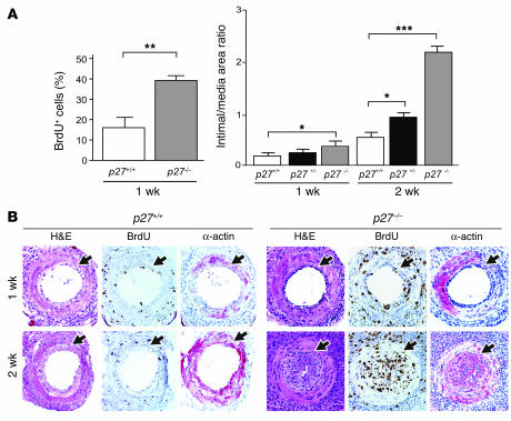
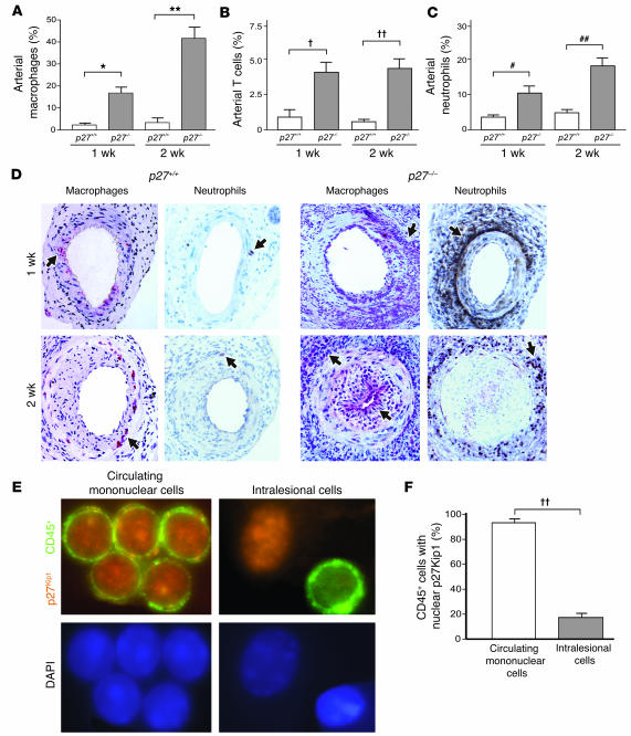
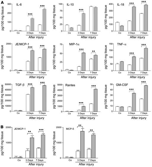
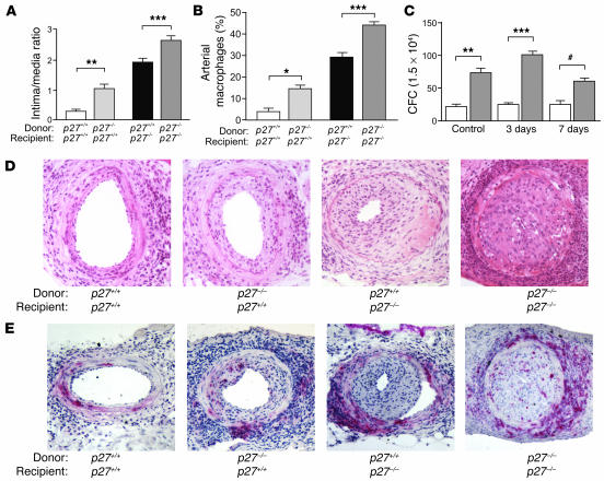
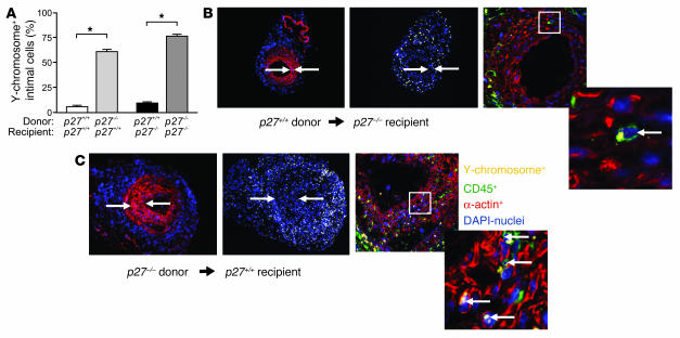
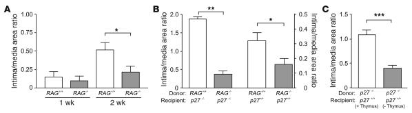
The cyclin-dependent kinase inhibitors are key regulators of cell cycle progression. Although implicated in carcinogenesis, they inhibit the proliferation of a variety of normal cell types, and their role in diverse human diseases is not fully understood. Here, we report that p27(Kip1) plays a major role in cardiovascular disease through its effects on the proliferation of bone marrow-derived (BM-derived) immune cells that migrate into vascular lesions. Lesion formation after mechanical arterial injury was markedly increased in mice with homozygous deletion of p27(Kip1), characterized by prominent vascular infiltration by immune and inflammatory cells. Vascular occlusion was substantially increased when BM-derived cells from p27(-/-) mice repopulated vascular lesions induced by mechanical injury in p27(+/+) recipients, in contrast to p27(+/+) BM donors. To determine the contribution of immune cells to vascular injury, transplantation was performed with BM derived from RAG(-/-) and RAG(+/+) mice. RAG(+/+) BM markedly exacerbated vascular proliferative lesions compared with what was found in RAG(-/-) donors. Taken together, these findings suggest that vascular repair and regeneration is regulated by the proliferation of BM-derived hematopoietic and nonhematopoietic cells through a p27(Kip1)-dependent mechanism and that immune cells largely mediate these effects.


Figures

Figure 1
Figure 1
Impaired wound healing in p27–/– mice in vivo. (A) Increased cell proliferation (left) and enlarged vascular lesions (right) in p27–/– arteries. *P < 0.05; **P < 0.01; ***P < 0.0001. (B) Representative cross sections of p27+/+ (left) and p27–/– (right) arteries stained with H&E, BrdU, and smooth muscle α-actin, 1 week and 2 weeks after injury. Arrows indicate the internal elastic lamina. Original magnification, ×200.
Figure 2
Figure 2
p27–/–mice develop arterial inflammation after vascular injury. (A_C) Accumulation of macrophages (*P< 0.0005; **P< 0.0001) (A), T lymphocytes ( P< 0.005; P< 0.001) (B), and neutrophils (#P< 0.01; ##P< 0.0005) (C) in p27+/+(white bars) and p27–/– (gray bars) arteries after injury. (D) Representative photomicrographs of cross sections of p27+/+ (left) and p27–/– (right) arteries immunostained for macrophages (arrow, red cytoplasmic staining) and neutrophils (arrow, brown cytoplasmic staining). Original magnification, ×200. (E) Immunofluorescence of circulating mononuclear (upper left) and intralesional cells (upper right) demonstrating coexpression of CD45 (membranes, green) and p27Kip1 (nuclei, red). Nuclear DAPI expression (blue) is also shown (lower panels). Original magnification, ×1000. (F) Quantitative analysis of coexpression of endogenous nuclear p27Kip1 in circulating CD45+ mononuclear cells (white bar) and intralesional cells (gray bar). Results are expressed as a percentage of CD45+, p27Kip1+ cells compared with CD45+ cells alone.
Figure 3
Figure 3
Cytokine levels are increased in p27–/– arteries following vascular injury. (A) Artery samples were collected from p27+/+ (white bars) and p27–/– (gray bars) mice at baseline (Co) and at 3 days and 7 days after injury. (B) Vascular tissues were extracted from p27+/+ (white bars) and p27–/– (gray bars) mice receiving p27+/+ BM at baseline and at 3 days and 7 days after injury. **P < 0.005; ***P < 0.0005.
Figure 4
Figure 4
p27Kip1 determines vascular proliferation and BM progenitor pool size. (A) p27–/– BM accelerates arterial lesion formation when transplanted into p27+/+ or p27–/– recipient mice. Following engraftment, arteries were injured and intima/media ratios were measured 2 weeks later. **P < 0.0005; ***P < 0.0001. (B) p27–/– BM significantly increased the percentage of arterial macrophages in p27+/+ and p27–/– recipient arteries compared with p27+/+ BM. *P < 0.005. (C) CFCs are significantly elevated in p27–/– BM (gray bars) compared with p27+/+ BM (white bars) at the indicated time points after vascular injury. Each data point was generated by three limiting dilutions. #P < 0.001. (D) Representative H&E-stained cross sections of recipient p27+/+ (left two panels) and p27–/– (right two panels) arteries following transplantation with donor p27+/+ and p27–/– BM, as indicated, followed by vascular injury. Original magnification, ×200. (E) Representative cross sections of recipient p27+/+ (left two panels) and p27–/– (right two panels) arteries immunostained for macrophages following transplantation with donor p27+/+ and p27–/– BM, as indicated, followed by vascular injury. Original magnification, ×200.
Figure 5
Figure 5
p27–/– BM–derived cells reconstitute the intima and adventitia of vascular lesions during repair. (A) The percentage of Y chromosome–positive nuclei in the intima was determined 2 weeks after injury using FISH techniques. *P < 0.0001. (B and C) Representative cross sections of recipient p27+/+ female arteries following transplantation of male p27+/+ (B) or p27–/– (C) donor BM. Left two panels: Y chromosome+ cells (yellow), α-actin+ cells (red), and nuclei (DAPI stain, blue). Arrows indicate the margins of the intima as determined by the internal and external elastic lamina. Right two panels: triple immunofluorescence detects Y chromosome+ cells (yellow), CD45+ cells (green), α-actin+ cells (red), and nuclei (DAPI stain, blue). Arrows indicate Y chromosome+ nuclei. Original magnification, ×100 (left two panels of B and C); right two panels: confocal microscopy.
Figure 6
Figure 6
Knockout of RAG confers protection against vascular proliferation. (A) RAG–/– arteries are protected against abnormal lesion formation 1 week and 2 weeks after vascular injury compared with RAG+/+ arteries. *P < 0.05. (B) RAG–/– BM directly reduces arterial lesion size in p27+/+ or p27–/– recipient mice. **P < 0.0001. (C) Thymectomy (T cell depletion) in p27+/+ mice reduces neointima development following transplantation with p27–/– BM. ***P < 0.0005.

References

    1. Ridker PM, Rifai N, Rose L, Burning JE, Cook NR. Comparison of C-reactive protein and low-density lipoprotein cholesterol levels in the prediction of first cardiovascular events. N. Engl. J. Med. 2002;347:1557–1565. - PubMed
    1. Binder CJ, et al. Innate and acquired immunity in atherogenesis. Nat. Med. 2002;8:1218–1226. - PubMed
    1. Libby P. Inflammation in atherosclerosis. Nature. 2002;420:868–874. - PubMed
    1. Hansson GK, Libby P, Schonbeck U, Yan ZQ. Innate and adaptive immunity in the pathogenesis of atherosclerosis. Circ. Res. 2002;91:281–291. - PubMed
    1. Polyak K, et al. Cloning of p27Kip1, a cyclin-dependent kinase inhibitor and a potential mediator of extracellular antimitogenic signals. Cell. 1994;78:59–66. - PubMed

Publication types

MeSH terms

Substances