Comparative Study
. 2004 Aug 1;18(15):1862-74.
doi: 10.1101/gad.1213904.

N-terminal polyubiquitination and degradation of the Arf tumor suppressor

Affiliations
Comparative Study

N-terminal polyubiquitination and degradation of the Arf tumor suppressor

Mei-Ling Kuo et al. Genes Dev. .

Abstract

Unknown mechanisms govern degradation of the p19Arf tumor suppressor, an activator of p53 and inhibitor of ribosomal RNA processing. Kinetic metabolic labeling of cells with [3H]-leucine indicated that p19Arf is a relatively stable protein (half-life approximately 6 h) whose degradation depends upon the ubiquitin-proteasome pathway. Although p19Arf binds to the Mdm2 E3 ubiquitin protein ligase to activate p53, neither of these molecules regulates p19Arf turnover. In contrast, the nucleolar protein nucleophosmin/B23, which binds to p19Arf with high stoichiometry, retards its turnover, and Arf mutants that do not efficiently associate with nucleophosmin/B23 are unstable and functionally impaired. Mouse p19Arf, although highly basic (22% arginine content), contains only a single lysine residue absent from human p14ARF, and substitution of arginine for lysine in mouse p19Arf had no effect on its rate of degradation. Mouse p19Arf (either wild-type or lacking lysine) and human p14ARF undergo N-terminal polyubiquitination, a process that has not as yet been documented in naturally occurring lysine-less proteins. Re-engineering of the p19Arf N terminus to provide consensus sequences for N-acetylation limited Arf ubiquitination and decelerated its turnover.


Figures
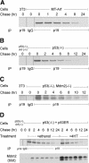
Figure 1.
Figure 1.
Kinetics of p19Arf turnover. (A) MT-Arf cells induced for 10 h with 100 μM ZnSO4 were starved for 30 min in fresh leucine-free medium, metabolically labeled for 2 h with10μCi/mL [3H]-leucine, and chased in fresh complete medium for the indicated times. Cell lysates were immunoprecipitated (IP) with antibodies to p19Arf or with control IgG as indicated below the panel. Lysates of control Arf-null NIH-3T3 cells treated similarly were also precipitated with antibodies to p19Arf (left lane). Proteins separated on denaturing gels were transferred to a membrane, treated with EN3HANCE, and subjected to radiofluorography at -80°C using an intensifying screen. (B) MEF cultures derived from p53-null embryos were starved of leucine, metabolically labeled with 100 μCi/mL [3H]-leucine, and the endogenously expressed Arf protein was analyzed as above. MEFs lacking both p53 and Arf were used as a control (left lane). (C) MEFs lacking p53 and Mdm2 were analyzed identically to those in B. (D) p53-null MEFs engineered to express p53-ER™ were starved and labeled with 100 μCi/mL [3H]-leucine. Cells were chased in fresh medium containing ethanol or 4-hydroxytamoxifen (4HT) as indicated, and lysates were precipitated with antibodies to p19Arf and analyzed (top panel). Aliquots of the same lysates were immunoblotted with antibodies to Mdm2 (bottom panel), a representative p53-inducible gene.
Figure 2.
Figure 2.
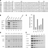
Analysis of Arf mutants. (A) N-terminal sequences encoded by Arf exon 1β are shown. (Hs) Homo sapiens; (Mm) Mus musculus; (Md) Monodelphis domestica, opossum; (Rn) Rattus norvegicus; (Ss) Sus scrofa, domestic pig; (Ma) Mesocricetus auratus, golden hamster; (Gg) Gallus gallus, chicken. (B) Lysates of NIH-3T3 cells infected with retroviruses encoding the indicated Arf mutants or an empty vector (Ctrl) were separated on denaturing gels, transferred to membrane, and blotted with antibodies to the indicated proteins. p53-null cells (lane 7) that overexpress endogenous p19Arf were used as a control, and antibodies to actin were used to control for protein loading. (C) NIH-3T3 cells infected as in B were stained with propidium iodide 48 h postinfection and analyzed for DNA content by flow cytometry. The percentage of cells in S phase was determined compared to the S-phase fraction (normalized to 100%) of cells infected with a control vector lacking Arf. (D) NIH-3T3 cells infected for 48 h with the indicated Arf or control retroviral vectors were metabolically labeled for 30 min with 2.5 μCi/mL [3H]-uridine and reincubated in fresh medium for 2 h. Where indicated, cells were exposed to 5 μM 5-fluorouridine (5-FU) 15 min prior to metabolic labeling to prevent rRNA processing. Extracted RNAs were separated on 1% agarose gels, transferred to a Hybond N+ membrane, treated with EN3HANCE, and subjected to autofluororadiography at -80°C using an intensifying screen. The positions of 47/45S and 32S rRNA precursors and mature 28S and 18S rRNAs are indicated. (E) NIH-3T3 cells infected with retroviral vectors encoding the indicated Arf mutants were subjected to pulse-chase analysis 48 h postinfection as in Figure 1A.
Figure 3.
Figure 3.
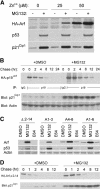
Stabilization of Arf by proteasome inhibition. (A) MT-Arf cells induced for 10 h with the indicated concentrations of ZnSO4 were cultured in fresh medium containing DMSO or 10 μM MG132 for an additional 8 h. Proteins from cell lysates were separated on a denaturing gel and blotted with antibodies to the indicated proteins. (B) Kinetic analysis of p19Arf turnover was performed as in Figure 1A, except that MT-Arf cells were induced with 50 μM ZnSO4, after which DMSO or MG132 was added to the labeling and chase medium. Proteins were blotted with antibodies to p21Cip1, which accumulates in response to MG132. Actin was used to control for protein loading. (C) NIH-3T3 cells infected for 48 h with retroviral vectors encoding the indicated Arf mutants were treated for an additional 24 h with DMSO, MG132, or E64 prior to lysis, electrophoresis, and immunoblotting with antibodies to the indicated proteins. Actin was used as a loading control. (D) Pulse-chase analysis was performed as in B using NIH-3T3 cells infected with a retroviral vector encoding HA-tagged Arf Δ2–14. Cell lysates were blotted with antibodies to p21Cip1 to demonstrate stabilization by MG132.
Figure 4.
Figure 4.
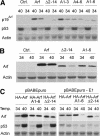
Turnover of Arf proteins in cells expressing temperature-sensitive E1 ubiquitin-activating enzyme. (A) Hamster ts-BN75 cells infected for 24 h with retroviruses packaged with VSV G protein and encoding the indicated Arf proteins were shifted to 34°C or 40°C for an additional 24 h. Cell lysates were separated on gels and blotted with antibodies to the indicated proteins. (B) NIH-3T3 cells infected with retroviral vectors encoding p19Arf and the indicated mutants were analyzed as in A. (C) tsBN75 cells were infected with pBABEpuro retroviral vectors (packaged in VSV G protein) either lacking or containing the gene encoding the E1 ubiquitin-activating enzyme. After selection for 48 h in puromycin, surviving cells were expanded and reinfected with VSV G protein-packaged vectors encoding the indicated Arf proteins. Twenty-four hours postinfection, cells were shifted to 34°C or 40°C for an additional 24 h. Cell lysates were separated, transferred to membrane, and blotted with antibodies to the indicated proteins. Actin was used as a loading control in each experiment.
Figure 5.
Figure 5.
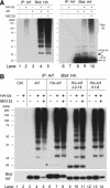
Mouse p19Arf is polyubiquitinated. (A) Human 293T cells were cotransfected with vectors encoding HA-tagged ubiquitin and/or p19Arf and, 24 h posttransfection, were either left untreated or were treated for 16 h with MG132. Lysates were immunoprecipitated (IP) with antibodies to p19Arf, and separated proteins were blotted with antibodies to the HA-tag (left) or to p19Arf (right). The mobilities of p19Arf, mono-ubiquitinated p19Arf (Arf-Ub), mono-HA-ubiquitinated p19Arf (Arf-HA-Ub), and polyubiquitinated forms (poly-Ub) are indicated at the far right. (B) Experiments analogous to those in A were performed using the indicated untagged or HA-tagged Arf proteins. The asterisk designates un-ubiquitinated HA-Arf.
Figure 6.
Figure 6.
E1 dependency and ubiquitination of human p14ARF and mouse lysine-less p19Arf. (A) ts-BN75 cells containing a thermolabile E1 enzyme were infected with a VSV G protein-packaged retroviral vector encoding untagged human p14ARF. Twenty-four hours postinfection, cells were shifted to 34°C or 40°C for an additional 24 h. Proteins separated on denaturing gels were transferred to membrane and blotted with antibodies to proteins indicated at the left. Actin was used as a loading control. (B) Human 293T cells transfected with retroviral expression vectors encoding untagged human p14ARF, human ARF Δ2–14, and/or HA-tagged ubiquitin (HA-Ub) were either left untreated or exposed to MG132. Equal amounts of lysate protein were precipitated with antibodies to human p14ARF, and separated proteins were blotted with antibodies to HA (top panel) or to p14ARF (bottom panel). (C) ts-BN75 cells infected with vectors encoding HA-tagged mouse p19Arf or a lysine-less mutant (K26R) were analyzed as in A. (D) Human 293T cells transfected with retroviral expression vectors encoding HA-tagged mouse p19Arf or the lysine-less K26R mutant were analyzed as in B. The asterisks in the upper panel indicate the position of the unubiquitinated HA-Arf proteins. (E) Pulsechase analyses of human p14ARF and p14ARF Δ2–14 (top panel) and mouse HA-p19Arf and the K26R mutant (bottom panel) expressed in NIH-3T3 cells.
Figure 7.
Figure 7.
N-terminal sequences determine Arf ubiquitination and turnover. (A) Human 293T cells transfected with HA-Arf Δ2–14 containing a factor Xa cleavage site C-terminal to the HA tag and/or His-ubiquitin were lysed and treated with protease Xa or not (indicated at top). Proteins recovered on nickel affinity columns in the presence of 8 M urea were separated on gels and blotted with anti-HA. The position of unmodified Arf protein is indicated by the arrow (left) and ubiquitinated species by asterisks. (Lanes 7,8) Longer exposures of lanes 5 and 6 reveal cleaved HA-tagged His-ubiquitin chains unbound to Arf. (B) 293T cells transfected with vectors encoding the indicated Arf N-terminal mutants and His-tagged ubiquitin were recovered, separated, and blotted with antibody to p19Arf. A background band at ∼55 kDa was detected in all lanes. Asterisks denote the lower-molecular-weight ubiquitinated Arf species. (Bottom) Immunoblotting was used to estimate the amounts of Arf protein loaded in each lane. (C) NIH-3T3 cells were infected with retroviral vectors encoding wild-type p19Arf and Arf variants engineered to undergo less efficient N-terminal ubiquitination. Pulse-chase analysis was used to compare the half-life of wild-type p19Arf (MGR, black circles) with the N-terminal mutants. (Gray circles) MDR; (black squares) MSA; (gray squares) MSR.
Figure 8.
Figure 8.
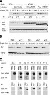
Stabilization of p19Arf by NPM/B23. (A) MT-Arf variants were derived by infection with a control retroviral vector expressing GFP or with vectors expressing Flag-tagged fulllength NPM or a truncated mutant (ΔC1) lacking the C-terminal RNA-binding domain. Cells induced for 10 h with 100 μM ZnS04 were incubated in fresh leucine-free medium for 30 min, labeled with [3H]-leucine for 2 h, and chased with complete medium for the indicated times. (Top) Cell lysates were immunoprecipitated (IP) with antibodies to p19Arf, and separated proteins were transferred to a membrane and detected by fluororadiography. (Bottom) Samples of the same cell lysates were separated and blotted with antibodies to the Flag-tag or to actin as a loading control. (B) MT-Arf cells infected with retroviruses encoding three different shRNAs to NPM/B23 were induced with 50 μM zinc for 10 h, after which zinc was removed. Cell lysates prepared at the indicated times thereafter (chase) were blotted with antibodies to p19Arf or NPM/B23. (C) 293T cells transfected with retroviral vectors encoding the indicated Arf proteins were lysed, and proteins (500 μg aliquots) were immunoprecipitated (IP) with normal mouse serum (NMS), normal rabbit serum (NRS), or antibodies to NPM or p19Arf. (Left) Separated proteins were transferred to a membrane and blotted with antibodies to NPM or Arf as indicated.

References

    1. Aviel S., Winberg, G., Massucci, M., and Ciechanover, A. 2000. Degradation of the Epstein-Barr virus latent membrane protein-1 (LMP1) by the ubiquitin–proteasome pathway. Targeting via ubiquitination of the N-terminal residue. J. Biol. Chem. 275: 23491-23499. - PubMed
    1. Bertwistle D., Sugimoto, M., and Sherr, C.J. 2004. Physical and functional interactions of the Arf tumor suppressor protein with nucleophosmin/B23. Mol. Cell. Biol. 24: 985-996. - PMC - PubMed
    1. Bloom J. and Pagano, M. 2004. To be or not to be.. .ubiquitinated. Cell Cycle 3: 138-140. - PubMed
    1. Bloom J., Amador, V., Bartolini, F., DeMartino, G., and Pagano, M. 2003. Proteasome-mediated degradation of p21 via N-terminal ubiquitinylation. Cell 115: 71-82. - PubMed
    1. Bradshaw R.A., Brickey, W.W., and Walker, K.W. 1998. N-terminal processing: The methionine aminopeptidase and Nα-acetyl transferase families. Trends Biochem. Sci. 23: 263-267. - PubMed

Publication types

MeSH terms