. 2004 Aug 2;166(3):407-18.
doi: 10.1083/jcb.200403003.

Selective modulation of type 1 insulin-like growth factor receptor signaling and functions by beta1 integrins

Affiliations

Selective modulation of type 1 insulin-like growth factor receptor signaling and functions by beta1 integrins

Hira Lal Goel et al. J Cell Biol. .

Abstract

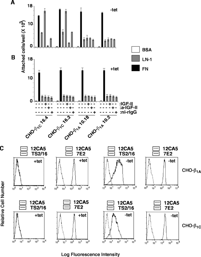
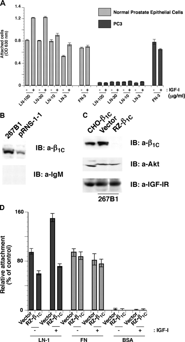
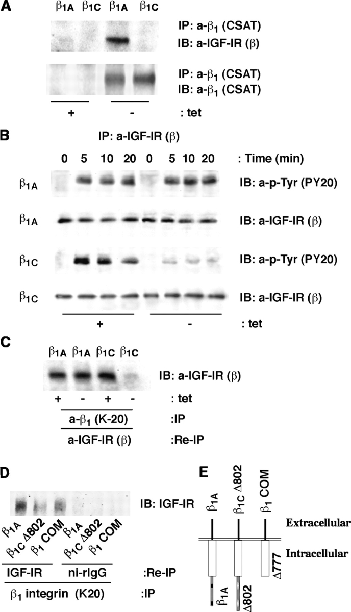
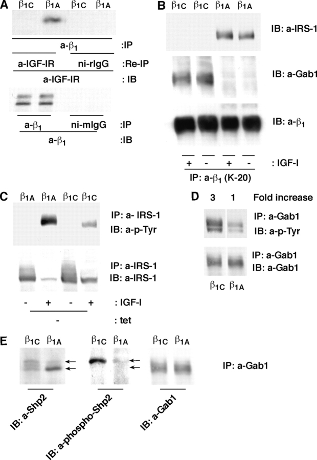
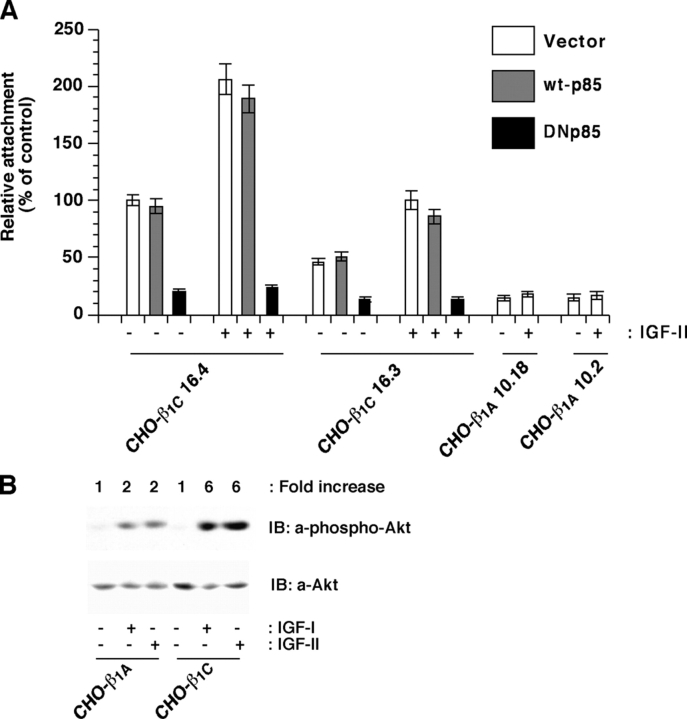
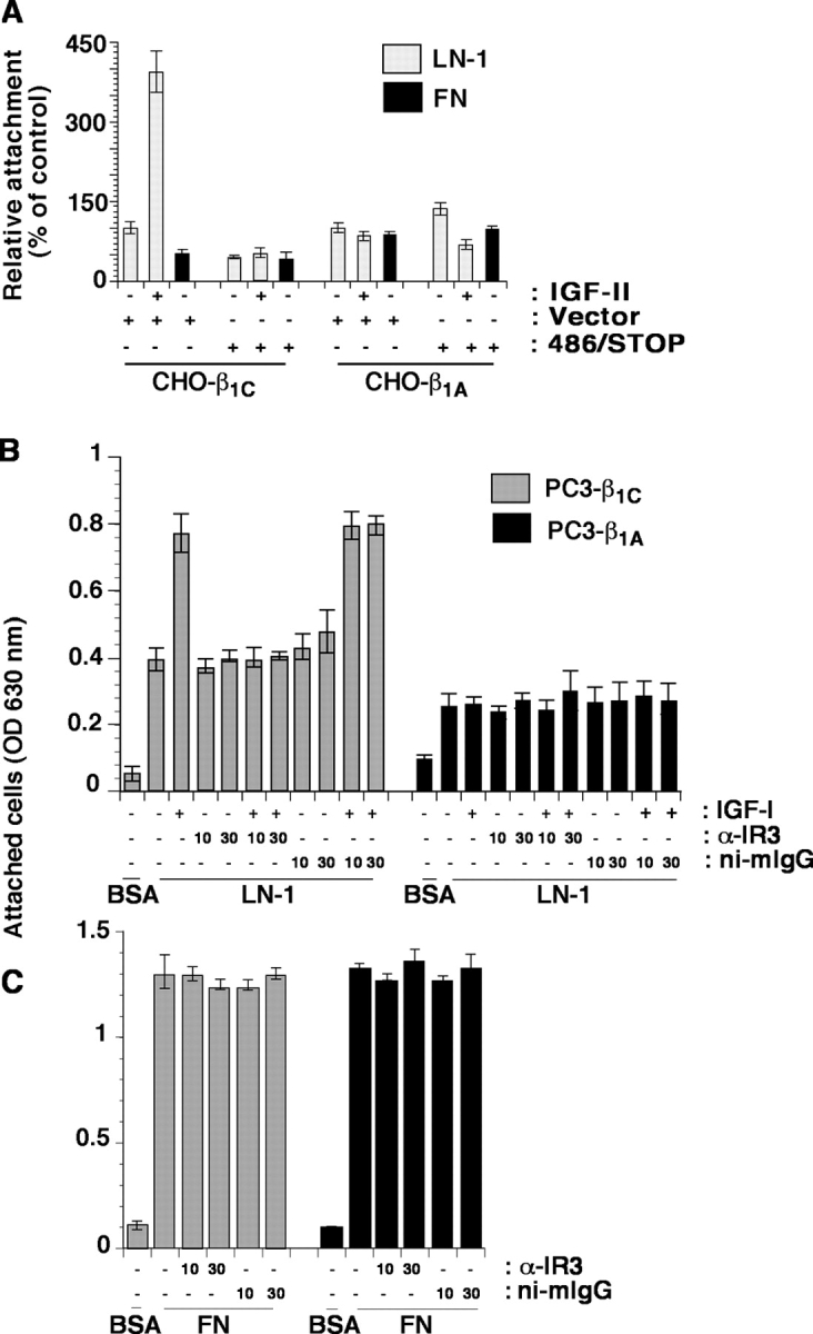
We show here that beta1 integrins selectively modulate insulin-like growth factor type I receptor (IGF-IR) signaling in response to IGF stimulation. The beta1A integrin forms a complex with the IGF-IR and insulin receptor substrate-1 (IRS-1); this complex does not promote IGF-I mediated cell adhesion to laminin (LN), although it does support IGF-mediated cell proliferation. In contrast, beta1C, an integrin cytoplasmic variant, increases cell adhesion to LN in response to IGF-I and its down-regulation by a ribozyme prevents IGF-mediated adhesion to LN. Moreover, beta1C completely prevents IGF-mediated cell proliferation and tumor growth by inhibiting IGF-IR auto-phosphorylation in response to IGF-I stimulation. Evidence is provided that the beta1 cytodomain plays an important role in mediating beta1 integrin association with either IRS-1 or Grb2-associated binder1 (Gab1)/SH2-containing protein-tyrosine phosphate 2 (Shp2), downstream effectors of IGF-IR: specifically, beta1A associates with IRS-1 and beta1C with Gab1/Shp2. This study unravels a novel mechanism mediated by the integrin cytoplasmic domain that differentially regulates cell adhesion to LN and cell proliferation in response to IGF.


Figures

Figure 1.
Figure 1.
IGF stimulates adhesion to LN-1 of β1C-expressing cells. β1C-CHO (clones 16.3 and 16.4) and β1A-CHO (clones 10.18 and 10.2) clones were cultured in the absence (A) or in the presence (B) of tet for 48 h. Cells were labeled using 51Cr-sodium chromate for 1 h at 37°C. 51Cr-labeled cells were incubated in the presence or in the absence of purified rabbit Ab to IGF-II or ni-rIgG for 1 h on ice. Cells were plated on BSA, LN-1, or FN at 37°C for 2 h in the presence or in the absence of IGF-II. Attached cells were washed, lysed, and the amount of 51Cr associated with the attached cells was measured by liquid scintillation counting. Data are expressed as mean ± SEM. (C) Representative β1C-CHO and β1A-CHO clones were cultured in the absence or in the presence of tet for 48 h and analyzed by FACS® using a β1 integrin Ab TS2/16 specific to human β1 or 7E2 specific to hamster β1 or, as negative control, 12CA5.
Figure 2.
Figure 2.
Down-regulation of β1C expression in 267B1 cells inhibits IGF-I–stimulated cell adhesion to LN-1. (A) PrEC and PC3 cells were serum starved for 12 h, detached, and plated on LN-1 or FN at 37°C for 2 h in the presence or in the absence of IGF-I. Cell adhesion was analyzed by crystal violet staining. (B) pRNS-1-1 and 267B1 cells were lysed and immunoblotted with mAb to β1C or normal IgM. (C) 267B1 cells were transiently transfected with pBJ1-RZ-β1C or vector alone, lysed, and immunoblotted with mAb to β1C, Ab to Akt, or Ab to IGF-IR. CHO-β1C cell lysate was used as a positive control. (D) 267B1 cells were transiently transfected with pBJ1-RZ-β1C or vector alone were detached and seeded on BSA or LN-1 or FN-coated plates at 37°C for 2 h in the presence or in the absence of IGF-I and stained with β-gal. Cell attachment was expressed as percentage of cells transfected with vector and attached to LN-1 in the absence of IGF-I, set at 100. Data are expressed as mean ± SEM. The experiments were repeated at least twice with similar results.
Figure 3.
Figure 3.
β1C enhances IGF-I– and II–mediated PC3 cell adhesion to LN-1. (A–D) β1A- and β1C-PC3 clones were cultured in the presence or in the absence of tet for 48 h. Cells were detached and plated on BSA or LN-1 or FN at 37°C for 2 h in the presence of IGF-II (A and B), of IGF-I (C and D), or of Ab to LN-1 (D) or ni-rIgG (D). In A, the differences in cell adhesion to LN-1 in the presence or in the absence of tet are statistically significant (*P ≤ 0.03). In C, the differences in cell adhesion to LN-1 between β1A and β1C expressing cells are statistically significant (*P ≤ 0.024). The experiments were repeated at least three times with similar results using two clones each of β1A- and β1C-PC3 cells. (E and F) β1A- and β1C-PC3 clones were cultured in the presence or in the absence of tet for 48 h. Cells were serum starved for 12 h, detached, and plated on LN-1 (E), FN or BSA (F) in serum-free medium at 37°C for 2 h in the presence or in the absence of IGF-I or GoH3 or rtIgG. Cell adhesion was analyzed by crystal violet staining. Data are expressed as mean ± SEM. (G) Surface expression of endogenous or exogenous β1A or β1C integrin or endogenous α6 integrin was analyzed in PC3 cells by FACS® using Ab to human β1, TS2/16, chicken β1, W1B10, α6, GoH3; or, as negative controls, mIgG, rtIgG, or 12CA5.
Figure 4.
Figure 4.
β1A and β1B differentially affect IGF-mediated cell proliferation and tumor growth. (A) β1A- and β1C-PC3 clones were cultured in the absence of tet for 48 h. Cells were plated on a 96-well plate in serum-free medium with or without IGF-I or IGF-II at 37°C. After 72 h of incubation, cell proliferation was analyzed using SRB. As control, sulforhodamine B (SRB) incorporation of cells attached to FN-coated substrates was measured in parallel (black bars). (B and C) PC3-β1A (B) and PC3-β1C (C) clones were injected subcutaneously in athymic male Balb/c mice. Mice were given water supplemented with either 5% sucrose to induce β1A or β1C expression, or 5% sucrose plus 100 μg/ml tet. The graphs show kinetics of tumor growth. Tumor growth is expressed as tumor volume in cubic millimeters. Data are the mean ± SEM of 10 animals per group. *P ≤ 0.001 at 10, 12, 14, and either 16 or 17 d from injection. These experiments were repeated three times with similar results.
Figure 5.
Figure 5.
IGF-IR and β1C act synergistically to support cell adhesion to LN-1. (A) R− and R+ cells were transiently transfected with human β1A or β1C. Surface expression of β1A or β1C was analyzed by FACS® using TS2/16 or 12CA5 as a negative control. Thick line, TS2/16; thin line, 12CA5. (B) R+ cells transiently transfected with β1A or β1C were detached and seeded on BSA or LN-1–coated plates at 37°C for 2 h in the presence or in the absence of IGF-II and stained with β-gal. (C) R− and R+ cells (106) transiently transfected with β1A or β1C were incubated with or without P4C10 (a-β1) or 1C10 (neg-cont) on ice for 1 h. Cells were plated on BSA or LN-1 or FN at 37°C for 2 h and stained with β-gal. Attachment of cells transfected with β1-integrin cDNA was expressed as percentage of cells transfected with pBJ (B) or pBJ-β1A (C) that were attached to LN-1, set at 100. The experiments were repeated at least twice with similar results. Data are expressed as mean ± SEM.
Figure 6.
Figure 6.
β1C inhibits IGF-IR tyrosine phosphorylation and β1A association with IGF-IR. β1A- and β1C-PC3 clones were cultured in the presence or in the absence of tet for 48 h, stimulated with IGF-I for 10 min at 37°C, lysed. Exogenous β1 was immunoprecipitated using CSAT (A) and endogenous β1 using K-20 (C). In A, immunoblotting (IB) was performed using CSAT or Ab to IGF-IR-β. (B) Cells were incubated with or without IGF-I for 0, 5, 10, and 20 min in suspension at 37°C. Immunoprecipitation was performed using Ab to IGF-IR-β. Immunoprecipitates were separated on SDS-PAGE and immunoblotted with PY20 or Ab to IGF-IR-β. In C, the endogenous β1 immunoprecipitates were reprecipitated and immunoblotted with Ab to IGF-IR-β. (D) CHO clones expressing the full-length β1A integrin or β1COM or β1CΔ802 truncated mutant were cultured for 48 h. Cells were lysed and proteins were immunoprecipitated with K-20. The immunocomplexes were dissociated and proteins were reimmunoprecipitated with Ab to IGF-IR or ni-rIgG. The immunoprecipitates were separated using SDS-PAGE and immunoblotted with Ab to IGF-IR. (E) Schematic representation of β1C cytoplasmic domain deletion mutants. The experiments were repeated at least twice with similar results.
Figure 7.
Figure 7.
β1A associates with and supports tyrosine phosphorylation of IRS-1, whereas β1C associates with and supports tyrosine phosphorylation of Gab1 and Shp2. (A) CHO clones expressing either β1C or β1A integrin were lysed and β1 integrin was immunoprecipitated using K-20. (A) The β1 immunoprecipitates were reprecipitated with Ab to IGF-IR or ni-rIgG and immunoblotted with polyclonal Ab to IGF-IR (top). As control, proteins from CHO cell lysates were immunoprecipitated with Ab to β1 integrin or ni-mIgG and immunoblotted with Ab to β1 integrin (Ab13, bottom). (B) CHO clones expressing either β1C or β1A integrin were incubated with or without IGF-I for 10 min, lysed, and proteins were immunoprecipitated with Ab to β1 integrins (K-20) and immunoblotted with Ab to IRS-1 (top), Gab1 (middle), or β1 integrin (bottom). (C–E) CHO clones expressing either β1C or β1A integrin were incubated with or without IGF-I for 10 min, lysed, and proteins were immunoprecipitated with Ab to IRS-1 (C) or Gab1 (D and E). Immunoprecipitates were separated using SDS-PAGE and immunoblotted with Ab to p-Tyr (PY20) (C and D), IRS-1 (C), Gab1 (D and E), Shp2 (E), or phospho-Shp2 (E). (F) β1A- and β1C-CHO clones were transiently transfected with wt-Shp2 or Shp2 C/S or vector alone. Cells were then cultured in the absence of tet for 48 h, plated on LN-1 and incubated for 2 h at 37°C in the presence or in the absence of IGF-I. After incubation, cells were fixed and stained with β-gal. Attachment of cells transfected with wt-Shp2 or Shp2 C/S cDNA was expressed as percentage of the number of attached cells transfected with vector alone, set at 100 in the absence of IGF-I. Data are expressed as mean ± SEM. (G) β1A- and β1C-CHO clones were transiently transfected with either HA-tagged wt-Shp2 or FLAG-tagged Shp2 C/S or vector alone. ERK1 was used as loading control. Cells were lysed after 48 h and immunoblotted with Ab to HA for wt-Shp2 or Ab to FLAG for Shp2 C/S. The experiments were repeated at least three times with similar results. White lines indicate that intervening lanes have been spliced out.
Figure 7.
Figure 7.
β1A associates with and supports tyrosine phosphorylation of IRS-1, whereas β1C associates with and supports tyrosine phosphorylation of Gab1 and Shp2. (A) CHO clones expressing either β1C or β1A integrin were lysed and β1 integrin was immunoprecipitated using K-20. (A) The β1 immunoprecipitates were reprecipitated with Ab to IGF-IR or ni-rIgG and immunoblotted with polyclonal Ab to IGF-IR (top). As control, proteins from CHO cell lysates were immunoprecipitated with Ab to β1 integrin or ni-mIgG and immunoblotted with Ab to β1 integrin (Ab13, bottom). (B) CHO clones expressing either β1C or β1A integrin were incubated with or without IGF-I for 10 min, lysed, and proteins were immunoprecipitated with Ab to β1 integrins (K-20) and immunoblotted with Ab to IRS-1 (top), Gab1 (middle), or β1 integrin (bottom). (C–E) CHO clones expressing either β1C or β1A integrin were incubated with or without IGF-I for 10 min, lysed, and proteins were immunoprecipitated with Ab to IRS-1 (C) or Gab1 (D and E). Immunoprecipitates were separated using SDS-PAGE and immunoblotted with Ab to p-Tyr (PY20) (C and D), IRS-1 (C), Gab1 (D and E), Shp2 (E), or phospho-Shp2 (E). (F) β1A- and β1C-CHO clones were transiently transfected with wt-Shp2 or Shp2 C/S or vector alone. Cells were then cultured in the absence of tet for 48 h, plated on LN-1 and incubated for 2 h at 37°C in the presence or in the absence of IGF-I. After incubation, cells were fixed and stained with β-gal. Attachment of cells transfected with wt-Shp2 or Shp2 C/S cDNA was expressed as percentage of the number of attached cells transfected with vector alone, set at 100 in the absence of IGF-I. Data are expressed as mean ± SEM. (G) β1A- and β1C-CHO clones were transiently transfected with either HA-tagged wt-Shp2 or FLAG-tagged Shp2 C/S or vector alone. ERK1 was used as loading control. Cells were lysed after 48 h and immunoblotted with Ab to HA for wt-Shp2 or Ab to FLAG for Shp2 C/S. The experiments were repeated at least three times with similar results. White lines indicate that intervening lanes have been spliced out.
Figure 8.
Figure 8.
PI 3-kinase mediates IGF-stimulated adhesion to LN-1. (A) β1C-CHO clones were transiently transfected with vector alone (pcDNA3), wt-p85, or DNp85. Cells were cultured in the absence of tet for 48 h. 1.5 × 10⁵ cells were plated per well coated with LN-1 and incubated for 2 h at 37°C in the presence or in the absence of 100 ng/ml IGF-II. Cells were washed, fixed, and stained with β-gal. Attachment of cells transfected with PI 3-kinase cDNA was expressed as percentage (average and SD) of the number of attached cells that were transfected with vector alone, set at 100. Data are expressed as mean ± SEM. (B) β1A- or β1C-CHO clones were stimulated with IGF-I or IGF-II for 10 min, lysed and immunoblotted with Ab to Akt or Ab to phospho-Akt. Experiments were repeated at least twice with similar results. Results using representative clones are shown.
Figure 9.
Figure 9.
IGF binding to IGF-IR mediates adhesion of β1C expressing cells to LN-1. (A) β1A- and β1C-CHO clones were transiently transfected with 486/STOP or vector alone. Cells were cultured in the absence of tet for 48 h. Cells were plated on either LN-1 or FN and incubated for 2 h at 37°C in the presence or in the absence of IGF-II. After incubation, cells were fixed and stained with β-gal. The number of attached cells transfected with 486/STOP cDNA was expressed as percentage of the number of attached cells transfected with vector alone, set at 100. (B and C) β1C- and β1A-PC3 clones were cultured in the absence of tet for 48 h. Cells were plated on BSA or LN-1 (B) or FN (C) at 37°C for 2 h in the presence or in the absence of IGF-I or α-IR3 or ni-mIgG. The experiments were repeated at least three times with similar results. Data are expressed as mean ± SEM.
Figure 10.
Figure 10.
A model for the biological effects of the cross-talk between IGF-IR and β1 integrins. This model shows that the β1C integrin forms a complex with and activates Gab1/Shp2; this results in recruitment of Shp2 to IGF-IR and consequently, IGF-IR dephosphorylation. The signaling events stimulated by β1C expression results in increased cell adhesion to laminin, reduced cell proliferation and inhibition of tumor growth. When down-regulation of β1C occurs, IGF-IR remains tyrosine phosphorylated and associated with β1A integrin, and this results in increased tumor growth and cell proliferation, but in reduced cell adhesion to LN.

Similar articles

Cited by

References

    1. Baserga, R. 2000. The contradictions of the insulin-like growth factor 1 receptor. Oncogene. 19:5574–5581. - PubMed
    1. Bello-DeOcampo, D., H.K. Kleinman, N.D. Deocampo, and M.M. Webber. 2001. Laminin-1 and α6β1 integrin regulate acinar morphogenesis of normal and malignant human prostate epithelial cells. Prostate. 46:142–153. - PubMed
    1. Bloch, W., E. Forsberg, S. Lentini, C. Brakebusch, K. Martin, H.W. Krell, U.H. Weidle, K. Addicks, and R. Fassler. 1997. β1 integrin is essential for teratoma growth and angiogenesis. J. Cell Biol. 139:265–278. - PMC - PubMed
    1. Bouvard, D., C. Brakebusch, E. Gustafsson, A. Aszodi, T. Bengtsson, A. Berna, and R. Fassler. 2001. Functional consequences of integrin gene mutations in mice. Circ. Res. 89:211–223. - PubMed
    1. Brar, P.K., B.L. Dalkin, C. Weyer, K. Sallam, I. Virtanen, and R.B. Nagle. 2003. Laminin α-1, α-3, and α-5 chain expression in human prepubertal benign prostate glands and adult benign and malignant prostate glands. Prostate. 55:65–70. - PubMed

Publication types