. 2004 Aug 2;200(3):367-76.
doi: 10.1084/jem.20040435.

Tumor necrosis factor (TNF) receptor shedding controls thresholds of innate immune activation that balance opposing TNF functions in infectious and inflammatory diseases

Affiliations

Tumor necrosis factor (TNF) receptor shedding controls thresholds of innate immune activation that balance opposing TNF functions in infectious and inflammatory diseases

Sofia Xanthoulea et al. J Exp Med. .

Abstract

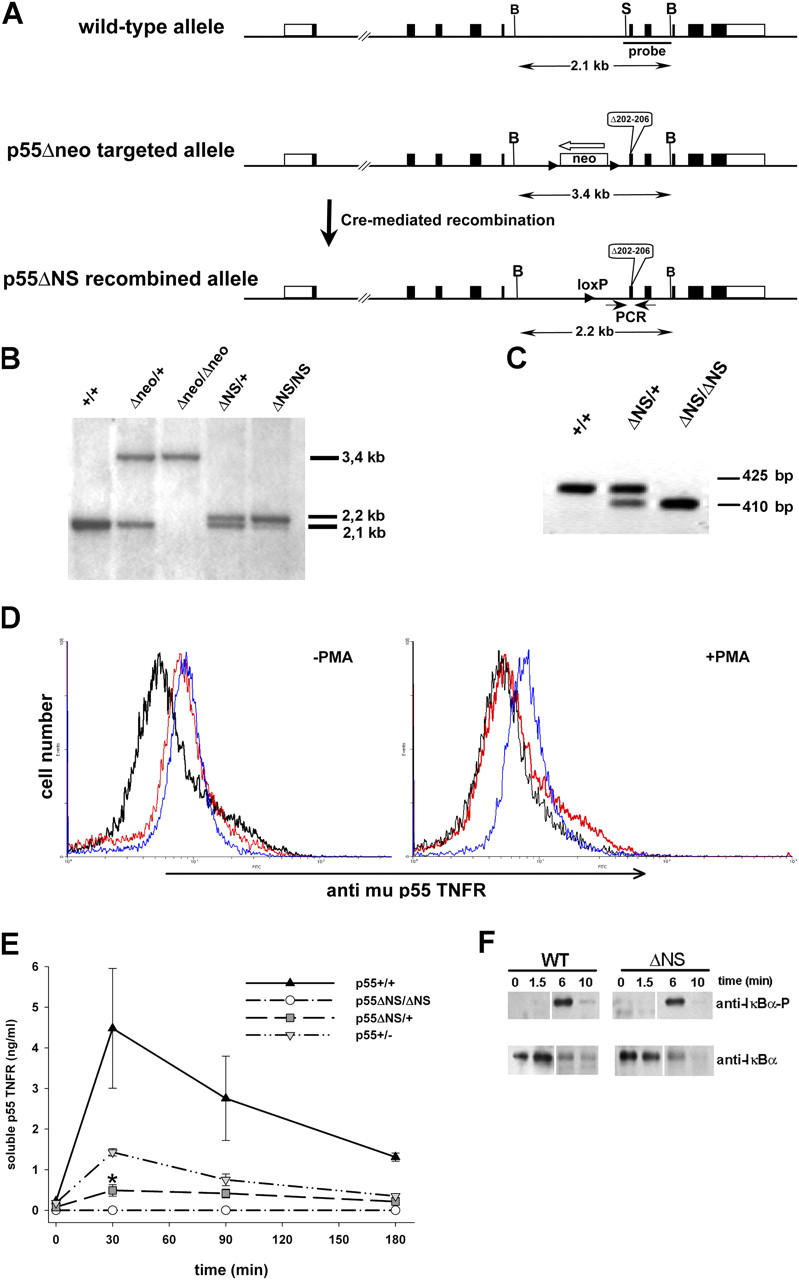
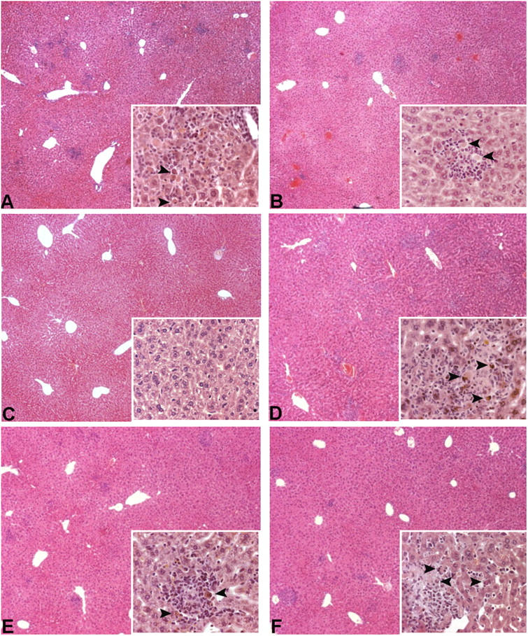
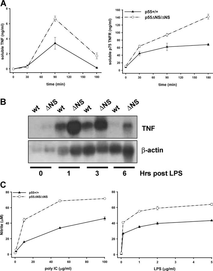
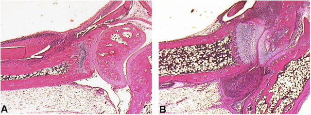
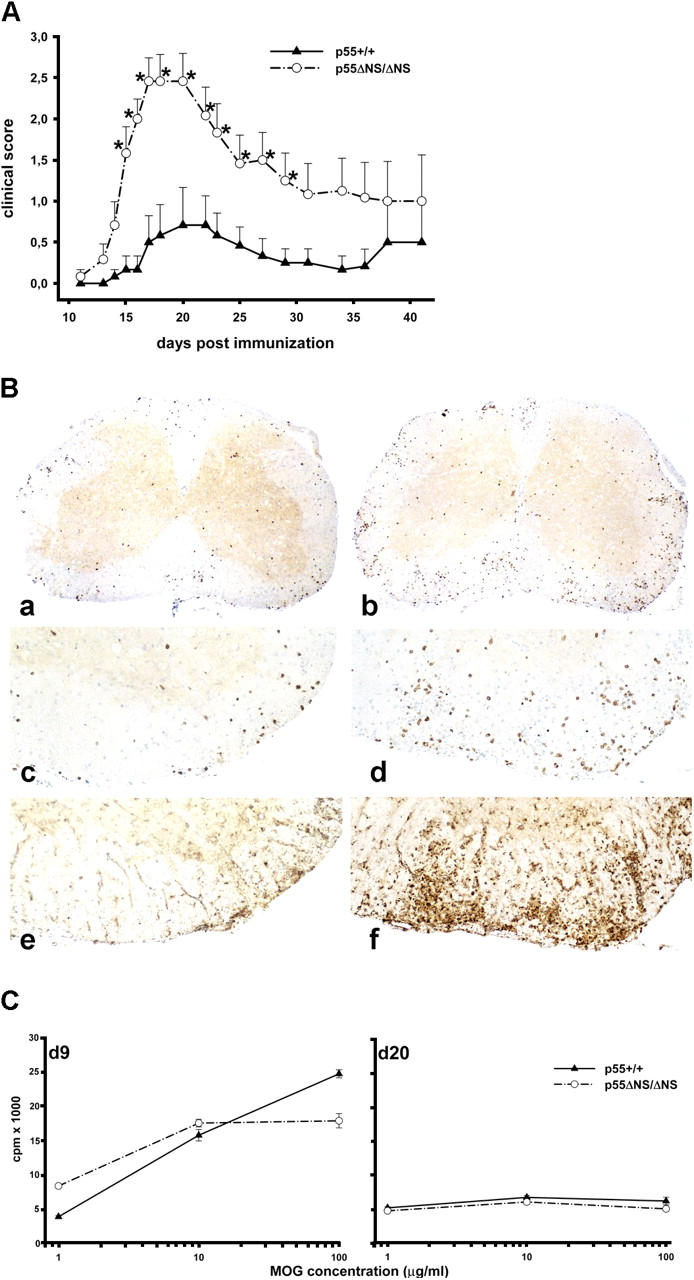
Tumor necrosis factor (TNF) is a potent cytokine exerting critical functions in the activation and regulation of immune and inflammatory responses. Due to its pleiotropic activities, the amplitude and duration of TNF function must be tightly regulated. One of the mechanisms that may have evolved to modulate TNF function is the proteolytic cleavage of its cell surface receptors. In humans, mutations affecting shedding of the p55TNF receptor (R) have been linked with the development of the TNFR-associated periodic syndromes, disorders characterized by recurrent fever attacks and localized inflammation. Here we show that knock-in mice expressing a mutated nonsheddable p55TNFR develop Toll-like receptor-dependent innate immune hyperreactivity, which renders their immune system more efficient at controlling intracellular bacterial infections. Notably, gain of function for antibacterial host defenses ensues at the cost of disbalanced inflammatory reactions that lead to pathology. Mutant mice exhibit spontaneous hepatitis, enhanced susceptibility to endotoxic shock, exacerbated TNF-dependent arthritis, and experimental autoimmune encephalomyelitis. These results introduce a new concept for receptor shedding as a mechanism setting up thresholds of cytokine function to balance resistance and susceptibility to disease. Assessment of p55TNFR shedding may thus be of prognostic value in infectious, inflammatory, and autoimmune diseases.


Figures

Figure 1.
Figure 1.
Generation and initial characterization of p55 ΔNS mice. (A) Schematic representation of the murine p55TNFR genomic locus (top), the p55 Δneo targeted locus generated by homologous recombination in embryonic stem cells (middle), and the p55 ΔNS locus generated after Cre-mediated excision of the loxP-flanked neo cassette (bottom). Exons are indicated as closed boxes with open boxes representing 5′ and 3′ untranslated regions. The p55 ΔNS mutation that deletes amino acids 202–206 in the mature protein is indicated. The white arrow shows the transcriptional orientation of the neo cassette and arrowheads indicate loxP sites. B and S indicate BamHI and SmaI restriction enzyme sites, respectively. (B) Southern blot analysis of BamHI-digested tail DNA from mice with the indicated genotypes using the SmaI-BamHI probe. (C) PCR analysis of tail DNA from mice with the indicated genotypes using PCR primers (small arrows in A) that amplify fragments of 425 and 410 bp for the wild-type and the mutant alleles, respectively. (D) Cell surface p55TNFR levels on peritoneal exudate cells from p55TNFR-deficient (black), wild-type (red), and homozygous p55 ΔNS/ΔNS mice (blue) analyzed for mp55TNFR expression before or after PMA activation. Data are representative of two independent experiments. (E) ELISA analysis of soluble p55TNFR levels in sera from mice with the indicated genotypes before and after LPS stimulation. Data are representative of three independent experiments. (*, P < 0.05). (F) Western blot analysis of IκBα phosphorylation and degradation in wild-type (wt) and homozygous p55 ΔNS/ΔNS (ΔNS) MEFs stimulated with human TNF and harvested at the indicated time points. All mice were in a B6,129 genetic background. White lines indicate that intervening lanes have been spliced out.
Figure 2.
Figure 2.
Active chronic hepatitis in p55 ΔNS mice. Histologic examination of liver sections from B6,129 p55 ΔNS mice at 8 wk in different experimental groups. (A and B) Focal inflammation in liver parenchyma characterized by infiltration of lymphocytes and polymorphonuclear cells and by the presence of apoptotic hepatocytes (inserts). Disease develops in homozygous p55 ΔNS/ΔNS (A) as well as in heterozygous p55 ΔNS/+ (B) mutant mice, albeit with reduced severity. (C) Liver sections from p55 ΔNS/ΔNS .TNF −/− mice present no signs of pathology. Inflammatory foci and presence of apoptotic hepatocytes persist in the liver of p55 ΔNS/ΔNS .RAG-1 −/− (D), p55 ΔNS/ΔNS .lpr/lpr (E), or p55 ΔNS/ΔNS .p75TNFR −/− (F) double mutant mice. Magnification, 40 and 400 for the insets.
Figure 3.
Figure 3.
Innate immune hyperresponsiveness in p55 ΔNS mice. (A) ELISA analysis of soluble TNF and p75TNFR levels in sera of wild-type (p55 +/+) and homozygous p55 ΔNS/ΔNS mice (n = 3 per group) before and after intraperitoneal injection of LPS. Data are representative of three independent experiments. (B) Northern blot analysis of TNF mRNA in thioglycollate-elicited peritoneal macrophages obtained from wild-type (p55 +/+) and p55 ΔNS/ΔNS mice before or after stimulation with LPS. The membrane was stripped and rehybridized with a β actin probe. (C) NO production in culture supernatants of wild-type (p55 +/+) and p55 ΔNS/ΔNS thioglycollate-elicited peritoneal macrophages in response to poly IC or LPS stimulation. All mice were in a B6,129 genetic background. Data are representative of three independent experiments.
Figure 4.
Figure 4.
Increased susceptibility of p55 ΔNS mice to chronic arthritis. Histologic examination of ankle joints from Tg197.p55 +/+ (A) and Tg197.p55 ΔNS/ΔNS (B) littermates at 4 wk of age. Increased formation of the inflammatory pannus and areas of cartilage and bone erosion are evident in B, whereas mild hyperplasia of the synovial membrane and onset of infiltration of polymorphonuclear and lymphocytic inflammatory cells in the synovial space is seen in A. All mice were littermates in a mixed CBA,B6,129 background.
Figure 5.
Figure 5.
Increased susceptibility of p55ΔNS mice to EAE. (A) EAE was induced in C57BL/6 p55 ΔNS/ΔNS and age-matched C57BL/6 controls (10–12 wk of age; n = 6 per group), by MOG p35–55 immunization as described in Materials and Methods. Data presented are means ± SEM of disease scores. *, P < 0.05 as determined by Student's t test. Data are representative of four separate experiments. (B) Immunohistochemistry from C57BL/6 controls (a, c, and e) and C57BL/6 p55 ΔNS/ΔNS (b, d, and f) mice. In both cases the animals with the highest inflammatory index and clinical signs were taken. (a and b) CD3 staining in spinal cord sections showing lymphocyte infiltration. Inflammation in the p55 ΔNS/ΔNS mouse is more prominent (magnification, 37). Higher magnifications (80) of panels a and b are shown in panels c and d, respectively. In addition to higher numbers of inflammatory T cells, the p55 ΔNS/ΔNS mouse (f; magnification, 80) also reveals more pronounced expression of mac-3+ macrophages than the wild-type control (e; magnification, 80). (C) Similar MOG p35–55–specific T cell responses in p55 ΔNS/ΔNS and p55 +/+ mice. T cell priming was measured in LN cells isolated from C57BL/6 p55 ΔNS/ΔNS and age-matched C57BL/6 control mice (7–11 wk of age) 9 d after immunization with MOG peptide as described in Materials and Methods. MOG-specific memory responses were examined in total spleen cells of C57BL/6 p55 ΔNS/ΔNS and age-matched C57BL/6 controls 20 d after EAE induction. Proliferation was determined by [3H]thymidine incorporation in T cell proliferation assays in which T cells were incubated with the indicated concentrations of MOG peptide. For each curve, two to three mice of each genotype were pooled together. One representative set of experiments out of three performed is shown.

References

    1. Smith, C.A., T. Farrah, and R.G. Goodwin. 1994. The TNF receptor superfamily of cellular and viral proteins: activation, costimulation, and death. Cell. 76:959–962. - PubMed
    1. Wallach, D., E.E. Varfolomeev, N.L. Malinin, Y.V. Goltsev, A.V. Kovalenko, and M.P. Boldin. 1999. Tumor necrosis factor receptor and Fas signaling mechanisms. Annu. Rev. Immunol. 17:331–367. - PubMed
    1. Wajant, H., K. Pfizenmaier, and P. Scheurich. 2003. Tumor necrosis factor signaling. Cell Death Differ. 10:45–65. - PubMed
    1. Pasparakis, M., S. Kousteni, J. Peschon, and G. Kollias. 2000. Tumor necrosis factor and the p55TNF receptor are required for optimal development of the marginal sinus and for migration of follicular dendritic cell precursors into splenic follicles. Cell. Immunol. 201:33–41. - PubMed
    1. Steinshamn, S., M.H. Bemelmans, L.J. van Tits, K. Bergh, W.A. Buurman, and A. Waage. 1996. TNF receptors in murine Candida albicans infection: evidence for an important role of TNF receptor p55 in antifungal defense. J. Immunol. 157:2155–2159. - PubMed

Publication types

MeSH terms

Substances