Marek's disease virus-encoded Meq gene is involved in transformation of lymphocytes but is dispensable for replication
- PMID: 15289599
- PMCID: PMC511057
- DOI: 10.1073/pnas.0404508101
Marek's disease virus-encoded Meq gene is involved in transformation of lymphocytes but is dispensable for replication
Abstract
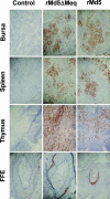
Marek's disease virus (MDV) causes an acute lymphoproliferative disease in chickens, resulting in T cell lymphomas in visceral organs and peripheral nerves. Earlier studies have determined that the repeat regions of oncogenic serotype 1 MDV encode a basic leucine zipper protein, Meq, which structurally resembles the Jun/Fos family of transcriptional activators. Meq is consistently expressed in MDV-induced tumor cells and has been suggested as the MDV-associated oncogene. To study the function of Meq, we have generated an rMd5DeltaMeq virus by deleting both copies of the meq gene from the genome of a very virulent strain of MDV. Growth curves in cultured fibroblasts indicated that Meq is dispensable for in vitro virus replication. In vivo replication in lymphoid organs and feather follicular epithelium was also not impaired, suggesting that Meq is dispensable for lytic infection in chickens. Reactivation of the rMd5DeltaMeq virus from peripheral blood lymphocytes was reduced, suggesting that Meq is involved but not essential for latency. Pathogenesis experiments showed that the rMd5DeltaMeq virus was fully attenuated in chickens because none of the infected chickens developed Marek's disease-associated lymphomas, suggesting that Meq is involved in lymphocyte transformation. A revertant virus that restored the expression of the meq gene, showed properties similar to those of the parental virus, confirming that Meq is involved in transformation but not in lytic replication in chickens.
Figures


Similar articles
-
Gene expression profiling in rMd5- and rMd5deltameq-infected chickens.Avian Dis. 2011 Sep;55(3):358-67. doi: 10.1637/9608-120610-Reg.1. Avian Dis. 2011. PMID: 22017031
-
Cell culture attenuation eliminates rMd5ΔMeq-induced bursal and thymic atrophy and renders the mutant virus as an effective and safe vaccine against Marek's disease.Vaccine. 2012 Jul 20;30(34):5151-8. doi: 10.1016/j.vaccine.2012.05.043. Epub 2012 Jun 8. Vaccine. 2012. PMID: 22687760
-
Deletion of the Meq gene significantly decreases immunosuppression in chickens caused by pathogenic Marek's disease virus.Virol J. 2011 Jan 5;8:2. doi: 10.1186/1743-422X-8-2. Virol J. 2011. PMID: 21205328 Free PMC article.
-
T-cell transformation by Marek's disease virus.Trends Microbiol. 1999 Jan;7(1):22-9. doi: 10.1016/s0966-842x(98)01427-9. Trends Microbiol. 1999. PMID: 10068994 Review.
-
Latency and tumorigenesis in Marek's disease.Avian Dis. 2013 Jun;57(2 Suppl):360-5. doi: 10.1637/10470-121712-Reg.1. Avian Dis. 2013. PMID: 23901747 Review.
Cited by
-
A multiplex xTAG assay for the simultaneous detection of five chicken immunosuppressive viruses.BMC Vet Res. 2018 Nov 15;14(1):347. doi: 10.1186/s12917-018-1663-1. BMC Vet Res. 2018. PMID: 30442149 Free PMC article.
-
Marek's Disease Virus Virulence Genes Encode Circular RNAs.J Virol. 2022 May 11;96(9):e0032122. doi: 10.1128/jvi.00321-22. Epub 2022 Apr 12. J Virol. 2022. PMID: 35412345 Free PMC article.
-
Targeted Editing of the pp38 Gene in Marek's Disease Virus-Transformed Cell Lines Using CRISPR/Cas9 System.Viruses. 2019 Apr 26;11(5):391. doi: 10.3390/v11050391. Viruses. 2019. PMID: 31027375 Free PMC article.
-
The Meq Genes of Nigerian Marek's Disease Virus (MDV) Field Isolates Contain Mutations Common to Both European and US High Virulence Strains.Viruses. 2024 Dec 31;17(1):56. doi: 10.3390/v17010056. Viruses. 2024. PMID: 39861844 Free PMC article.
-
Marek's disease viral interleukin-8 promotes lymphoma formation through targeted recruitment of B cells and CD4+ CD25+ T cells.J Virol. 2012 Aug;86(16):8536-45. doi: 10.1128/JVI.00556-12. Epub 2012 May 30. J Virol. 2012. PMID: 22647701 Free PMC article.
References
-
- Calnek, B. W. (2001) Curr. Top. Microbiol. Immunol. 255, 25–55. - PubMed
-
- Peng, Q., Zeng, M., Bhuiyan, Z. A., Ubukata, E., Tanaka, A., Nonoyama, M. & Shirazi, Y. (1995) Virology 213, 590–599. - PubMed
-
- Ross, N. L. (1999) Trends Microbiol. 7, 22–29. - PubMed
-
- Ross, N., O'Sullivan, G., Rothwell, C., Smith, G., Burgess, S. C., Rennie, M., Lee, L. F. & Davison, T. F. (1997) J. Gen. Virol. 78, 2191–2198. - PubMed
Publication types
MeSH terms
Substances
LinkOut - more resources
Full Text Sources
Other Literature Sources
Miscellaneous

