

Genes encoding candidate pheromone receptors in a moth (Heliothis virescens)
- PMID: 15289611
- PMCID: PMC511062
- DOI: 10.1073/pnas.0403052101
Genes encoding candidate pheromone receptors in a moth (Heliothis virescens)
Abstract
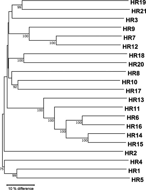
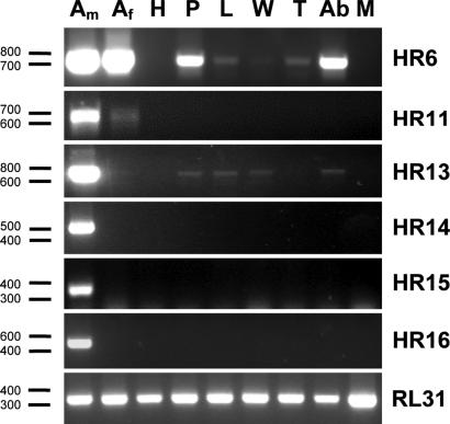
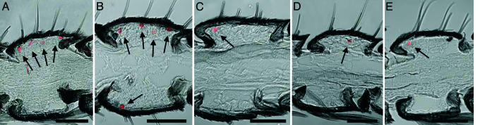
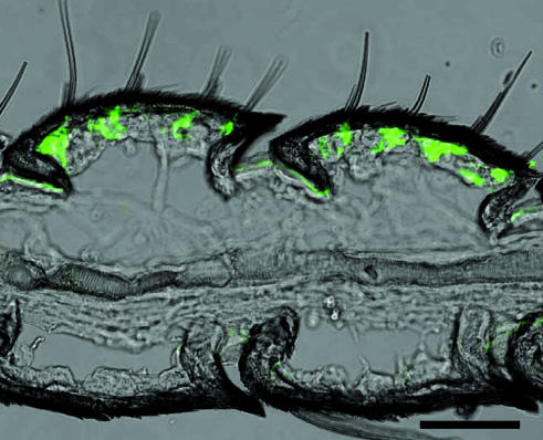
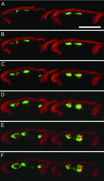
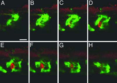
The remarkable responsiveness of male moths to female released pheromones is based on the extremely sensitive and selective reaction of highly specialized sensory cells in the male antennae. These cells are supposed to be equipped with male-specific receptors for pheromonal compounds, however, the nature of these receptors is still elusive. By using a combination of genomic sequence analysis and cDNA-library screening, we have cloned various cDNAs of the tobacco budworm Heliothis virescens encoding candidate olfactory receptors. A comparison of all identified receptor types not only highlighted their overall high degree of sequence diversity but also led to the identification of a small group of receptors sharing >40% identity. In RT-PCR analysis it was found that distinct members of this group were expressed exclusively in the antennae of male moths. In situ hybridization experiments revealed that the male-specific expression of these receptor types was confined to antennal cells located beneath sensillar hair structures (sensilla triochoidea), which have been shown to contain pheromone-sensitive neurons. Moreover, two-color double in situ-hybridization approaches uncovered that cells expressing one of these receptor types were surrounded by cells expressing pheromone-binding proteins, as expected for a pheromone-sensitive sensillum. These findings suggest that receptors like Heliothis receptor 14-16 (HR14-HR16) may render antennal cells responsive to pheromones.
Figures
Similar articles
-
Candidate pheromone receptors provide the basis for the response of distinct antennal neurons to pheromonal compounds.Eur J Neurosci. 2007 Apr;25(8):2364-73. doi: 10.1111/j.1460-9568.2007.05512.x. Eur J Neurosci. 2007. PMID: 17445234
-
Larval sensilla of the moth Heliothis virescens respond to sex pheromone components.Insect Mol Biol. 2016 Oct;25(5):666-78. doi: 10.1111/imb.12253. Epub 2016 Jul 28. Insect Mol Biol. 2016. PMID: 27465144
-
Molecular elements of pheromone detection in the female moth, Heliothis virescens.Insect Sci. 2018 Jun;25(3):389-400. doi: 10.1111/1744-7917.12434. Epub 2017 Mar 1. Insect Sci. 2018. PMID: 28026117
-
Working range of stimulus flux transduction determines dendrite size and relative number of pheromone component receptor neurons in moths.Chem Senses. 2012 May;37(4):299-313. doi: 10.1093/chemse/bjr122. Epub 2012 Jan 9. Chem Senses. 2012. PMID: 22230170 Review.
-
The multiple role of the pheromone-binding protein in olfactory transduction.Ciba Found Symp. 1996;200:267-75; discussion 275-80. doi: 10.1002/9780470514948.ch19. Ciba Found Symp. 1996. PMID: 8894303 Review.
Cited by
-
Analysis of the Antennal Transcriptome and Identification of Tissue-specific Expression of Olfactory-related Genes in Micromelalopha troglodyta (Lepidoptera: Notodontidae).J Insect Sci. 2022 Sep 1;22(5):8. doi: 10.1093/jisesa/ieac056. J Insect Sci. 2022. PMID: 36165424 Free PMC article.
-
Characterization of an enantioselective odorant receptor in the yellow fever mosquito Aedes aegypti.PLoS One. 2009 Sep 15;4(9):e7032. doi: 10.1371/journal.pone.0007032. PLoS One. 2009. PMID: 19753115 Free PMC article.
-
A family of chemoreceptors in Tribolium castaneum (Tenebrionidae: Coleoptera).PLoS One. 2007 Dec 19;2(12):e1319. doi: 10.1371/journal.pone.0001319. PLoS One. 2007. PMID: 18091992 Free PMC article.
-
Odorant Receptor PxylOR11 Mediates Repellency of Plutella xylostella to Aromatic Volatiles.Front Physiol. 2022 Jul 13;13:938555. doi: 10.3389/fphys.2022.938555. eCollection 2022. Front Physiol. 2022. PMID: 35910574 Free PMC article.
-
Antennal transcriptome of Manduca sexta.Proc Natl Acad Sci U S A. 2011 May 3;108(18):7449-54. doi: 10.1073/pnas.1017963108. Epub 2011 Apr 15. Proc Natl Acad Sci U S A. 2011. PMID: 21498690 Free PMC article.
References
-
- Hildebrand, J. G. & Shepherd, G. M. (1997) Annu. Rev. Neurosci. 20, 595-631. - PubMed
-
- Steinbrecht, R. A. (1997) Int. J. Insect Morphol. Embryol. 26, 229-245.
-
- Krieger, J. & Breer, H. (1999) Science 286, 720-723. - PubMed
-
- Gao, Q. & Chess, A. (1999) Genomics 60, 31-39. - PubMed
-
- Clyne, P. J., Warr, C. G., Freeman, M. R., Lessing, D., Kim, J. & Carlson, J. R. (1999) Neuron 22, 327-338. - PubMed
Publication types
MeSH terms
Substances
Associated data
- Actions
- Actions
- Actions
- Actions
- Actions
- Actions
- Actions
- Actions
- Actions
- Actions
- Actions
- Actions
LinkOut - more resources
Full Text Sources
