. 2004 Aug;186(16):5486-95.
doi: 10.1128/JB.186.16.5486-5495.2004.

Identification of the secretion and translocation domain of the enteropathogenic and enterohemorrhagic Escherichia coli effector Cif, using TEM-1 beta-lactamase as a new fluorescence-based reporter

Affiliations

Identification of the secretion and translocation domain of the enteropathogenic and enterohemorrhagic Escherichia coli effector Cif, using TEM-1 beta-lactamase as a new fluorescence-based reporter

Xavier Charpentier et al. J Bacteriol. 2004 Aug.

Abstract

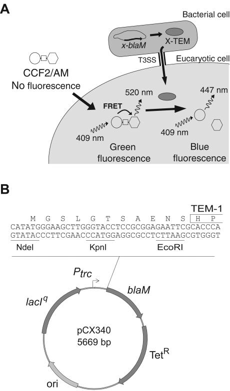
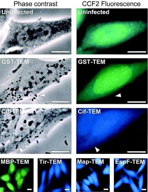
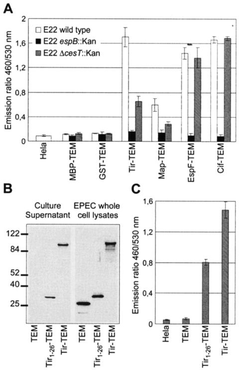
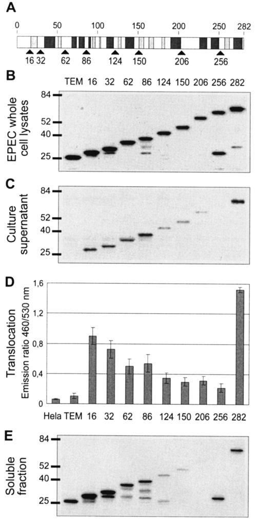
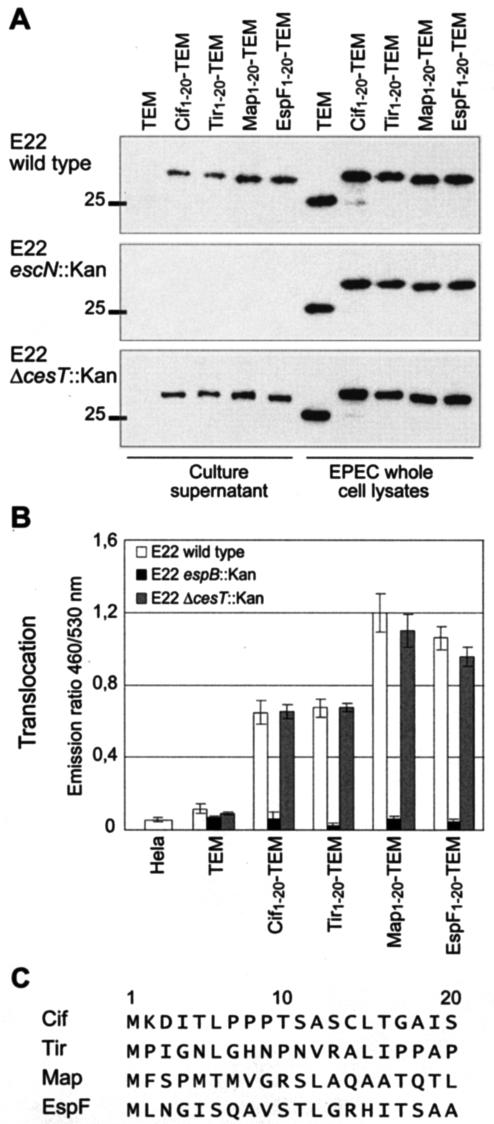
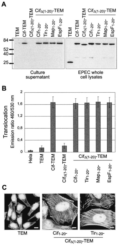
Enteropathogenic and enterohemorrhagic Escherichia coli (EPEC and EHEC) strains are human and animal pathogens that inject effector proteins into host cells via a type III secretion system (TTSS). Cif is an effector protein which induces host cell cycle arrest and reorganization of the actin cytoskeleton. Cif is encoded by a lambdoid prophage present in most of the EPEC and EHEC strains. In this study, we analyzed the domain that targets Cif to the TTSS by using a new reporter system based on a translational fusion of the effector proteins with mature TEM-1 beta-lactamase. Translocation was detected directly in living host cells by using the fluorescent beta-lactamase substrate CCF2/AM. We show that the first 16 amino acids (aa) of Cif were necessary and sufficient to mediate translocation into the host cells. Similarly, the first 20 aa of the effector proteins Map, EspF, and Tir, which are encoded in the same region as the TTSS, mediated secretion and translocation in a type III-dependent but chaperone-independent manner. A truncated form of Cif lacking its first 20 aa was no longer secreted and translocated, but fusion with the first 20 aa of Tir, Map, or EspF restored both secretion and translocation. In addition, the chimeric proteins were fully able to trigger host cell cycle arrest and stress fiber formation. In conclusion, our results demonstrate that Cif is composed of a C-terminal effector domain and an exchangeable N-terminal translocation signal and that the TEM-1 reporter system is a convenient tool for the study of the translocation of toxins or effector proteins into host cells.


Figures

FIG. 1.
FIG. 1.
Schematic representation of the TEM-1 reporter system used to study translocation of TTSS effectors into live eukaryotic cells. (A) Upon passive entry into the eukaryotic cell, the nonfluorescent esterified CCF2/AM substrate is rapidly converted by cellular esterases in charged and fluorescent CCF2. Excitation of the coumarin moiety (represented by a circle) at 409 nm results in fluorescence energy transfer (FRET) to the fluorescein moiety (represented by a hexagon), which emits a green fluorescence signal at 520 nm. Injection of an effector fused to TEM-1 into a CCF2-loaded cell induces catalytic cleavage of the CCF2 β-lactam ring (represented by a square), disrupting FRET. This produces an easily detectable and measurable change in CCF2 fluorescence from green to blue emission. (B) Map of the effector-TEM fusion cloning vector. The blaM gene encodes the mature form of the β-lactamase TEM (the first two residues are boxed). The NdeI, KpnI, and EcoRI restriction sites are unique. The origin of replication (ori) is derived from ColE1.
FIG. 2.
FIG. 2.
Demonstration of the translocation of EPEC effector proteins into live HeLa cells by using TEM-1 fusions and fluorescence microscopy. HeLa cells were infected with wild-type EPEC strains expressing different TEM-1 fusion proteins. After infection, HeLa cells were washed and loaded with CCF2/AM. β-Lactamase activity in HeLa cells is revealed by the blue fluorescence emitted by the cleaved CCF2 product, whereas uncleaved CCF2 emits a green fluorescence. No detectable fluorescence arises from adherent bacteria (indicated by arrowheads). Bars, 10 μM.
FIG. 3.
FIG. 3.
Analysis of the role of the translocator, chaperone, and STS with the TEM-1 translocation reporter system. (A) Activity of the EspB translocator and the CesT chaperone on effector translocation. HeLa cells were infected with wild-type EPEC E22 strains expressing MBP-TEM or GST-TEM fusion protein or the different effector-TEM protein fusions. After infection, HeLa cells were washed and loaded with CCF2/AM. β-Lactamase activity in HeLa cells was detected by measuring cleavage of the CCF2/AM substrate with a fluorescence microplate reader and is presented as the emission ratio between blue fluorescence (460 nm) and green fluorescence (530 nm). (B) Secretion of TEM-1 fused to the secretion signal of Tir. Culture supernatants from wild-type E22 expressing TEM-1 alone, the Tir1-26-TEM fusion, and the Tir-TEM fusion were subjected to Western blot analysis with anti-TEM-1 antibody. Molecular mass markers (in kilodaltons) are indicated to the left. (C) Translocation of TEM-1 fused to the secretion signal of Tir. HeLa cells were infected with E22 strains expressing TEM-1 alone, the Tir1-26-TEM fusion, and the Tir-TEM fusion. The presented translocation data are averages of triplicate values of the results from three experiments.
FIG. 4.
FIG. 4.
Identification of the minimal N-terminal domain of Cif that can mediate translocation into eukaryotic cells. (A) Schematic representation of the predicted secondary structure of Cif. Light and dark gray boxes represent β-sheets and α-helices, respectively. Black arrows indicate the different sites of fusion to TEM-1. (B) Expression of Cif1-X-TEM fusions in EPEC. EPEC whole-cell lysates were subjected to Western blot analysis with anti-TEM-1 antibody. Molecular mass markers (in kilodaltons) are indicated to the left. (C) Secretion of the produced Cif1-X-TEM fusions. Culture supernatants from E22 Δcif expressing TEM-1 alone and Cif1-X-TEM fusions were subjected to Western blot analysis with anti-TEM-1 antibody. (D) Translocation of Cif1-X-TEM fusions in HeLa cells. The presented data are averages of triplicate values of the results from three experiments. Numbers indicate the different sites of Cif fused to TEM-1. (E) Solubility analysis of the produced Cif1-X-TEM fusions. EPEC cells were lysed, and the soluble fraction was obtained after the removal of insoluble proteins, cell debris, and unbroken cells by centrifugation.
FIG. 5.
FIG. 5.
The first 20 residues of Cif, Tir, Map, and EspF mediate both secretion and translocation of TEM-1 in a type III-dependent manner. (A) Production and secretion of Cif1-20-TEM, Tir1-20-TEM, Map1-20-TEM, and EspF1-20-TEM fusions in wild-type E22, the escN mutant (TTSS defective), and the cesT mutant (defective for Tir/Map chaperone). Molecular mass markers (in kilodaltons) are indicated to the left. (B) Translocation in HeLa cells of Cif1-20-TEM, Tir1-20-TEM, Map1-20-TEM, and EspF1-20-TEM fusions in wild-type E22, the cesT mutant, and the espB mutant (translocation defective). The presented data are averages of triplicate values of the results from three experiments. (C) Alignment of the first 20 residues of Cif, Tir, Map, and EspF.
FIG. 6.
FIG. 6.
Cif is composed of a C-terminal effector domain and an exchangeable N-terminal translocation signal. (A) Production and secretion of chimeric Cif-TEM fusions. Molecular mass markers (in kilodaltons) are indicated to the left. (B) Translocation in HeLa cells of chimeric Cif-TEM fusions. The presented data are averages of triplicate values of the results from two independent experiments. (C) CPE triggered by chimeric Cif-TEM fusions. HeLa cells were infected under conditions used to monitor translocation. At the end of the interaction, bacteria were killed with gentamicin and HeLa cells were incubated for a further 3 days. Actin and nuclei were stained, respectively, with rhodamine-phalloidin and DAPI. Bars, 10 μM.

References

    1. Abe, A., M. de Grado, R. A. Pfuetzner, C. Sanchez-Sanmartin, R. Devinney, J. L. Puente, N. C. Strynadka, and B. B. Finlay. 1999. Enteropathogenic Escherichia coli translocated intimin receptor, Tir, requires a specific chaperone for stable secretion. Mol. Microbiol. 33:1162-1175. - PubMed
    1. Charpentier, X., C. Chalut, M. H. Remy, and J. M. Masson. 2002. Penicillin-binding proteins 1a and 1b form independent dimers in Escherichia coli. J. Bacteriol. 184:3749-3752. - PMC - PubMed
    1. Combet, C., C. Blanchet, C. Geourjon, and G. Deleage. 2000. NPS@: network protein sequence analysis. Trends Biochem. Sci. 25:147-150. - PubMed
    1. Crawford, J. A., and J. B. Kaper. 2002. The N-terminus of enteropathogenic Escherichia coli (EPEC) Tir mediates transport across bacterial and eukaryotic cell membranes. Mol. Microbiol. 46:855-868. - PubMed
    1. Creasey, E. A., R. M. Delahay, A. A. Bishop, R. K. Shaw, B. Kenny, S. Knutton, and G. Frankel. 2003. CesT is a bivalent enteropathogenic Escherichia coli chaperone required for translocation of both Tir and Map. Mol. Microbiol. 47:209-221. - PubMed

Publication types

MeSH terms

LinkOut - more resources