. 2004 Oct;15(10):4669-81.
doi: 10.1091/mbc.e04-03-0265. Epub 2004 Aug 3.

Localization of RNAs to the mitochondrial cloud in Xenopus oocytes through entrapment and association with endoplasmic reticulum

Affiliations

Localization of RNAs to the mitochondrial cloud in Xenopus oocytes through entrapment and association with endoplasmic reticulum

Patrick Chang et al. Mol Biol Cell. 2004 Oct.

Abstract

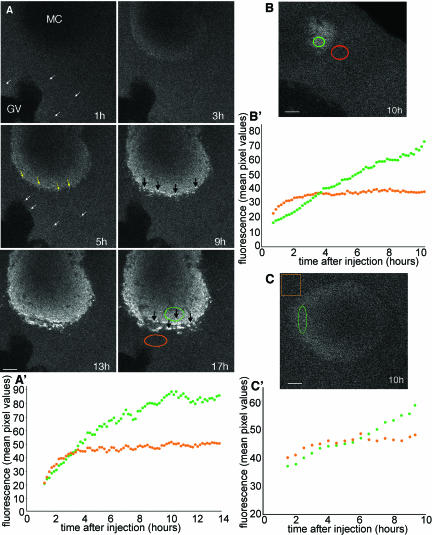
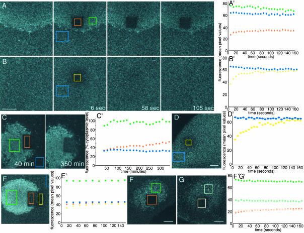
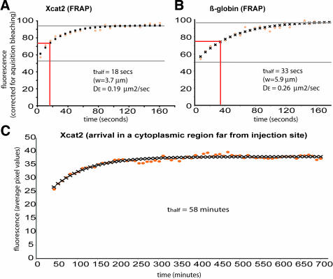
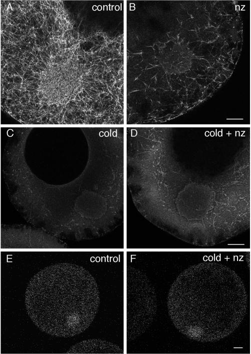
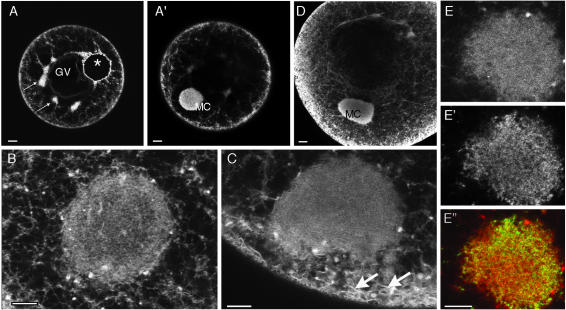
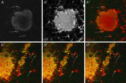
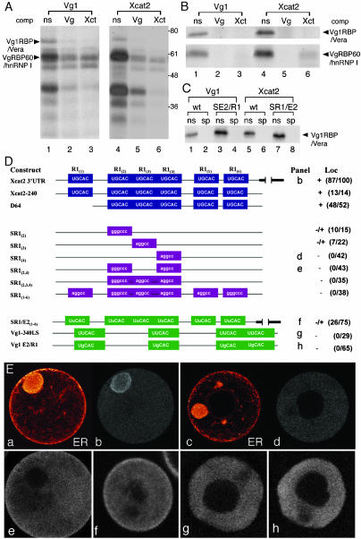
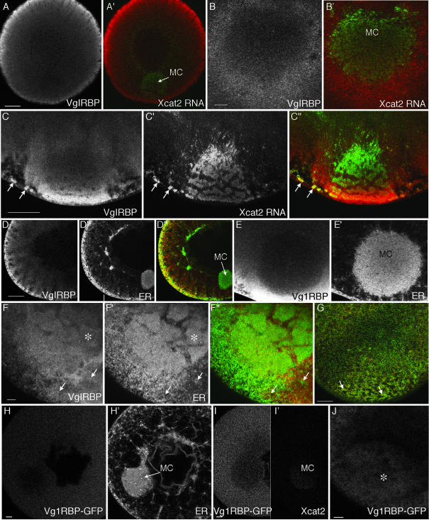
The germ cell lineage in Xenopus is specified by the inheritance of germ plasm, which originates within a distinct "mitochondrial cloud" (MC) in previtellogenic oocytes. Germ plasm contains localized RNAs implicated in germ cell development, including Xcat2 and Xdazl. To understand the mechanism of the early pathway through which RNAs localize to the MC, we applied live confocal imaging and photobleaching analysis to oocytes microinjected with fluorescent Xcat2 and Xdazl RNA constructs. These RNAs dispersed evenly throughout the cytoplasm through diffusion and then became progressively immobilized and formed aggregates in the MC. Entrapment in the MC was not prevented by microtubule disruption and did not require localization to germinal granules. Immobilized RNA constructs codistributed and showed coordinated movement with densely packed endoplasmic reticulum (ER) concentrated in the MC, as revealed with Dil16(3) labeling and immunofluorescence analysis. Vg1RBP/Vera protein, which has been implicated in linking late pathway RNAs to vegetal ER, was shown to bind specifically both wild-type Xcat2 3' untranslated region and localization-defective constructs. We found endogenous Vg1RBP/Vera and Vg1RBP/Vera-green fluorescent protein to be largely excluded from the MC but subsequently to codistribute with Xcat2 and ER at the vegetal cortex. We conclude that germ line RNAs localize into the MC through a diffusion/entrapment mechanism involving Vg1RBP/Vera-independent association with ER.


Figures

Figure 1.
Figure 1.
Progressive accumulation of Xcat2 3′UTR in the MC. (A) Confocal images from series acquired every 10 min during a 17-h period after microinjection of Alexa 488-Xcat2 RNA into a stage I oocyte (video 1). The particulate appearance of the fluorescent probe in the cytoplasm (white arrows) and the subsequent concentration of small particles in peripheral regions of the MC (yellow arrows), before the accumulation of progressively larger aggregates (black arrows) in peripheral regions of the MC, should be noted. (A′) Quantification of Xcat2 fluorescence in a peripheral region of the MC (green) and a nearby cytoplasmic region (orange). In the cytoplasmic region, fluorescence initially increased for a period of 2–3 h as it spread from the injection site (on the far side of the nucleus) and then remained at a constant level. In the MC, accumulation of Xcat2 3′UTR was linear for the first 5 h after injection. (B and B′) Quantification of fluorescent Xcat2 3′UTR accumulation in a peripheral region of the MC (green) (MC seen in tangential section) and a nearby cytoplasmic region (orange) in another stage I oocyte. (C and C′) Quantification of fluorescent Xdazl LE accumulation in a peripheral region of the MC (green) and a nearby cytoplasmic region (orange) in another stage I oocyte. All scale bars, 10 μm.
Figure 2.
Figure 2.
Immobilization of Xcat2 RNA in the MC. (A) Confocal images from a FRAP experiment performed on a stage I oocyte 20 h after injection of Alexa 488-Xcat2 3′UTR. Throughout the figure, unbleached regions of the MC are shown in green, unbleached regions of the cytoplasm in blue, bleached regions of the MC in orange, and bleached regions of the cytoplasm in yellow. Times after photobleaching are indicated. (A′) Quantification of fluorescence recovery in the bleached zone (orange), compared with nearby regions of the MC (green) and cytoplasm (blue). No transfer of fluorescence to the photobleached zone from surrounding regions was detected, indicating that Xcat2 3′UTR had become immobilized in the MC. (B and B′) In a cytoplasmic region (yellow) of the same oocyte, fluorescence recovered to the level of an unbleached region (blue) within 2 min after photobleaching. (C and C′) Long-term FRAP analysis of another oocyte injected with fluorescent Xcat2 3′UTR revealed ongoing linear RNA accumulation in the bleached zone of the MC (orange), indicating that photobleaching did not damage this region. The frontier of bleached material with the MC remained largely unaltered after several hours, as did unbleached MC (green) and cytoplasm (blue). (D and D′) FRAP analysis of a fluorescent β-globin RNA 3′UTR construct, which served as a nonlocalized control RNA, in a cytoplasmic region (yellow) of a stage I oocyte. Fluorescence recovery to the level of an unbleached cytoplasmic region (blue) was complete within 3 min. (E and E′) FRAP experiment with another oocyte injected with fluorescent Xcat2 3′UTR, showing fluorescence recovery to cytoplasmic levels both inside (orange) and outside (yellow) the MC during the 3 min after bleaching but no redistribution of localized fluorescent RNA within the MC (see video 2). (F, F′, G, and G′) Localized fluorescent Xdazl RNA (F and F′) (dark orange/dark green) behaved similarly to Xcat2 3′UTR (A and A′) after photobleaching, as did Xcat2 3′UTR lacking the GGLE (G and G′) (light orange/light green) in another oocyte, showing that RNA immobilization does not require incorporation into germinal granules (see video 3). In each case, the y axis is given in arbitrary units of fluorescence as mean pixel values within the region shown. All scale bars, 10 μm.
Figure 3.
Figure 3.
Diffusion of injected fluorescent RNAs in oocyte cytoplasm. Diffusion of fluorescent Xcat2 3′UTR (A and C) and the β-globin RNA 3′UTR construct (B) injected into stage I oocytes and incubated overnight, as in Figures 1 and 2, was analyzed. FRAP was monitored in A and B, and the arrival of fluorescence in a cytoplasmic region far from the injection site was monitored in C. Theoretical diffusion curves (black crosses) were found to fit closely to the experimental data (orange circles), plotted as mean pixel values in a cytoplasmic region within the bleached zone, normalized relative to an unbleached zone at each time point in the FRAP experiments. Effective diffusion constants (DE) were calculated from the FRAP data (see Materials and Methods).
Figure 4.
Figure 4.
Microtubule-independent Xcat2 localization. (A–D) Confocal images of oocytes fixed for antitubulin immunofluorescence after treatments to disrupt microtubules. In oocytes cultured in control medium for 24 h, microtubules were detected as a dense network both inside and outside the MC (A). After 24 h of culture at 18°C with 10 μM nocodazole (∼30 μg/ml), a significant population of microtubules remained (B). In contrast, a 90-min cold treatment destroyed almost all detectable microtubules (C), and regrowth was prevented with culture of such oocytes at 18°C for 24 h with 10 μM nocodazole (D). (E and F) Fluorescent Xcat2 3′UTR in oocytes cultured at 18°C in control medium (E) and with nocodazole after 90-min cold treatment (F) localized to the MC to equivalent degrees (groups of 6 or 7 were examined). Scale bars, 25 μm.
Figure 5.
Figure 5.
The MC contains a dense ER network. (A—C) Confocal images of stage I (A and B) and stage II (C) oocytes injected with oil droplets (*) loaded with the lipophilic dye DiI. The dye spread into a network extending throughout the cell (A), was enriched in cortical regions as well as in small clumps around the GV (small arrows), and was highly concentrated in the MC, lying in a different focal plane (A′). At higher magnification, a dense network could be discriminated within the MC in some oocytes (B). In stage II oocytes (C), ER sheets in the subcortical region (arrows) appeared to merge with ER in the MC. (D) Immunofluorescence assays with an antibody to the ER luminal protein GRP94 revealed a distribution very similar to that of DiI staining, as seen in this stage II oocyte. (E, E′, and E″) Comparison of labeling with DiI (E) (at high magnification, in an optical plane through the periphery of a MC) after oil droplet injection the previous day with that with the short-chain permeable dye DiO (E′). As seen in the overlay (E″, DiI in red and DiO in green), the two dyes labeled clearly distinct membranous organelles, probably mainly ER (DiI) and mitochondria (DiO). Scale bars, 20 μm in A and D, 10 μm in B, C, and E.
Figure 6.
Figure 6.
Localized RNAs associate with ER. (A) Confocal images of a live stage I oocyte injected the previous day with Alexa 488-Xcat2 3′UTR (A) and DiI-loaded oil drop (A′). RNA aggregates (green in the overlay A″) are observed to coincide in positions in and around the MC (e.g., at arrows) and with accumulations of ER (red in A″). (B) Higher magnification, consecutive, overlaid images (colors as in A″) from a sequence acquired every 6.5 s in a confocal plane tangential to the edge of the MC, showing that Xcat2 particles (green) undergo jostling movements together with the ER (red) (e.g., at arrows; see also video 4).
Figure 7.
Figure 7.
Vg1RBP/Vera binding to early pathway RNA. (A) The Vg1 LS and Xcat2 3′UTR bind to similar oocyte factors. Radiolabeled Vg1 LS (lanes 1–3) and Xcat2 3′UTR (lanes 4–6) transcripts were tested with UV cross-linking for the ability to be bound in vitro by proteins from an S10 oocyte lysate. Specificity of binding was assessed by competition with unlabeled non-specific competitor RNA (ns, lanes 1 and 4), Vg1 LS (Vg, lanes 2 and 5), or Xcat2 3′UTR (Xct, lanes 3 and 6). Vg1RBP/Vera and VgRBP60/hnRNP I are indicated on the left; molecular weight markers are indicated on the right. (B) Both Vg1RBP/Vera and VgRBP60/hnRNP I specifically bind to the Xcat2 3′UTR. Radiolabeled Vg1 LS (lanes 1–3) and Xcat2 3′UTR (lanes 4–6) transcripts were tested by UV cross-linking for the ability to be bound in vitro by partially purified Vg1RBP/Vera (top) and VgRBP60/hnRNP I (bottom). Specificity of binding was assessed by competition with unlabeled nonspecific competitor RNA (ns, lanes 1 and 4), Vg1 LS (Vg, lanes 2 and 5), or Xcat2 3′UTR (Xct, lanes 3 and 6). (C) Mutations within E2 or R1 motifs do not affect binding of Vg1RBP/Vera to the Vg1 LS and Xcat2 3′UTR. Radiolabeled Vg1 LS (wt, lanes 1 and 2), Vg1 E2/R1 (SE2/R1, lanes 3 and 4), Xcat2 3′UTR (wt, lanes 5 and 6), or SR1/E2(1–6) (SR1/E2, lanes 7 and 8) transcripts were tested with UV cross-linking for the ability to be bound in vitro by partially purified Vg1RBP/Vera. Specificity of binding was assessed by competition with unlabeled nonspecific competitor RNA (ns, odd-numbered lanes) or Vg1 LS (sp, even-numbered lanes). (D) UGCAC motifs are required for Xcat2 MC entrapment. The schematic representation of the 3′UTR of Xcat2 shows the UGCAC (R1) motifs and deletion (D) or noncognate substitution (S) mutants tested by microinjection into stage I oocytes. Numbers identify which R1 repeat is changed in the Xcat2 3′UTR. Point mutations changed R1 to E2 repeats in Xcat2 [SR1/E2(1–6)] or E2 to R1 repeats in the Vg1–340 LS (Vg1 E2/R1). Scores for localization (Loc) (right) indicate the number of oocytes showing localization per the total number of injected oocytes in which a MC was detected. Data were compiled from three independent experiments. Letters preceding the scores refer to panels in E. (E) Confocal images showing localization of representative oocytes injected with selected Alexa 488-labeled transcripts diagramed in D. Panels a and c show the ER network labeled with DiI of the oocytes shown in b and d, respectively. Oocyte diameters 150–300 μm.
Figure 8.
Figure 8.
Vg1RBP/Vera is excluded from the MC but enriched in the vegetal cortex. (A–G) Confocal images showing immunofluorescence of stage I (A, B, D, and E), early stage II (C and F), and stage III (G) oocytes with an anti-Vg1RBP/Vera antibody. At all stages, staining was observed throughout the cytoplasm, being enriched in cortical regions and relatively weak in the MC. (A′, B′, and C″) Superimposed images with anti-Vg1RBP/Vera (red) and Alexa 488-labeled Xcat2 3′UTR injected 20 h before fixation (green; also shown in C′). Anti-Vg1RBP/Vera and localized fluorescent RNAs (arrows in C″) are found together in stage II oocytes around the MC and in the region of MC-cortex contact but not elsewhere. (D′, E′, and F′) Costaining with anti-GRP94 to visualize ER. (D″, F″, and G) Superimposed images, with anti-Vg1RBP/Vera in red and anti-GRP94 in green. Again, localization in the same region is observed only once the MC meets the vegetal cortex (F and F″), remaining colocalized in patches at the vegetal cortex in stage III (G). (H–J) Confocal images of live oocytes, 24 h after injection of RNA coding for Vg1RBP/Vera-GFP (H, I, and J) and either DiI (H′) or rhodamine-Xcat2 3′UTR (I′). Vg1RBP/Vera-EGFP protein was distributed homogeneously in stage I oocytes (H and I), with relatively little in the MC. It did not codistribute with either ER or exogenous localized Xcat2 3′UTR. In stage II (J), like exogenous Vg1RBP/Vera, it became concentrated at the vegetal pole (*). Scale bars, 10 μm in B, E, F, H, I, and J, 50 μm in A, C, D, and G.

References

    1. Allison, R, Czaplinski, K., Git, A., Adegbenro, E., Stennard, F., Houliston E., and Standart, N. (2004). Xenopus Staufen 1 and 2 were vegetally localized during oogenesis. RNA (in press). - PMC - PubMed
    1. Ainger, K., Avossa, D., Diana, A.S., Barry, C., Barbarese, E., and Carson, J.H. (1997). Transport and localization elements in myelin basic protein mRNA. J. Cell Biol. 138, 1077-1087. - PMC - PubMed
    1. Alarcon, V.B., and Elinson, R.P. (2001). RNA anchoring in the vegetal cortex of the Xenopus oocyte. J. Cell Sci. 114, 1731-1741. - PubMed
    1. Argon, Y., and Simen, B.B. (1999). GRP94, an ER chaperone with protein and peptide binding properties. Semin. Cell Dev. Biol. 10, 495-505. - PubMed
    1. Bashirullah, A., Cooperstock, R.L., and Lipshitz, H.D. (1998). RNA localization in development. Annu. Rev. Biochem. 67, 335-394. - PubMed

Publication types

LinkOut - more resources