. 2004 Oct;15(10):4500-11.
doi: 10.1091/mbc.e04-05-0432. Epub 2004 Aug 3.

Elevated endosomal cholesterol levels in Niemann-Pick cells inhibit rab4 and perturb membrane recycling

Affiliations

Elevated endosomal cholesterol levels in Niemann-Pick cells inhibit rab4 and perturb membrane recycling

Amit Choudhury et al. Mol Biol Cell. 2004 Oct.

Abstract

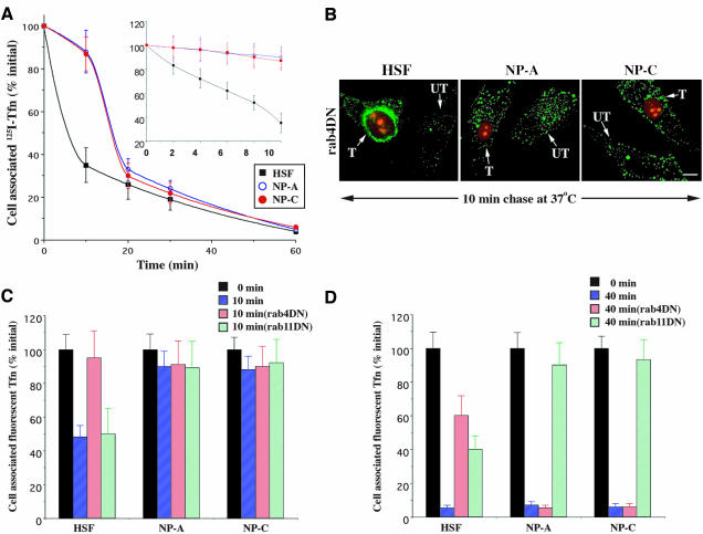
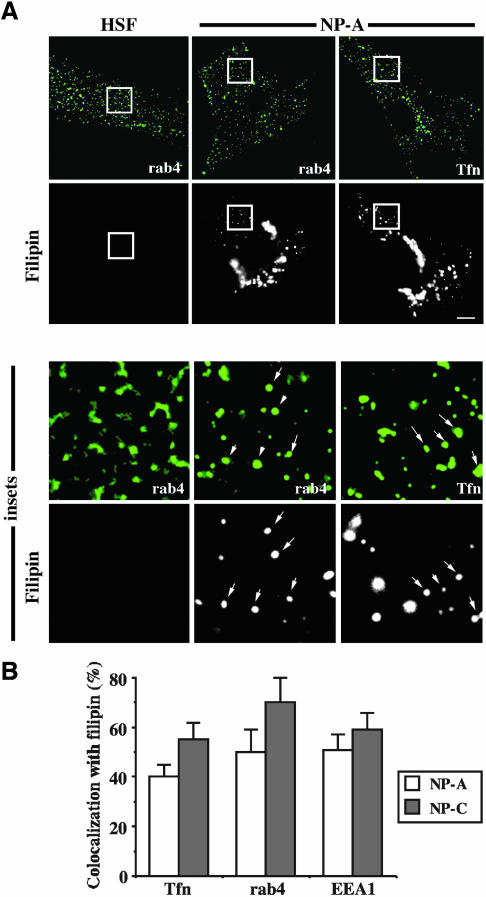
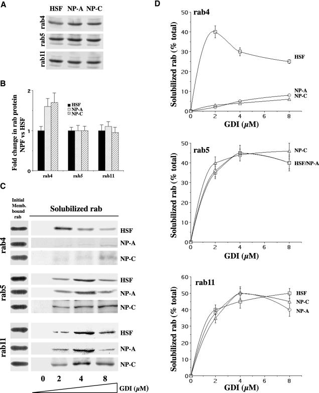
In normal human skin fibroblasts (HSFs), fluorescent glycosphingolipid analogues are endocytosed and sorted into two pools, one that is recycled to the plasma membrane and one that is transported to the Golgi complex. Here, we investigated glycosphingolipid recycling in Niemann-Pick type A and C lipid storage disease fibroblasts (NPFs). Cells were incubated with a fluorescent analogue of lactosylceramide (LacCer) at 16 degrees C to label early endosomes (EEs), shifted to 37 degrees C, and lipid recycling was quantified. Using dominant negative rabs, we showed that, in normal HSFs, LacCer recycling was rapid (t1/2 approximately 8 min) and mainly rab4-dependent. In NPFs, LacCer recycling was delayed (t1/2 approximately 30-40 min), and rab4-dependent recycling was absent, whereas rab11-dependent recycling predominated. Transferrin recycling via the rab4 pathway was similarly perturbed in NPFs. Compared with normal HSFs, EEs in NPFs showed high cholesterol levels and an altered organization of rab4. In vitro extraction of rab4 (but not rab11) with GDP dissociation inhibitor was severely attenuated in NPF endosomal fractions. This impairment was reversed with cholesterol depletion of isolated endosomes or with high-salt treatment of endosomes. These data suggest that abnormal membrane recycling in NPFs results from specific inhibition of rab4 function by excess cholesterol in EEs.


Figures

Figure 1.
Figure 1.
Altered recycling of BODIPY-LacCer in NPFs vs. normal HSFs. (A) Cells were incubated with 2.5 μM BODIPY-LacCer for 1 h at 16°C to selectively label EEs, washed, and back-exchanged with 5% DF-BSA to remove any fluorescent lipid remaining at the PM. The samples were then incubated for 0, 10, or 40 min at 37°C in the presence of 5% DF-BSA to remove any fluorescent LacCer that was recycled back to the PM. G, Golgi apparatus. Bar, 10 μm. (B) Cells were incubated with fluorescent LacCer as in A, and the amount of cell-associated fluorescence was determined by lipid extraction and analysis. Inset, the first 10 min of LacCer recycling was examined in detail. In some experiments (filled symbols), cells were grown in lipoprotein-deficient serum to deplete cholesterol (see Materials and Methods). All values are expressed as percentage of cell-associated fluorescence at 0 min for each cell type and are mean ± SD of three or more independent experiments.
Figure 2.
Figure 2.
Effects of DN rab4, DN rab11, and a PI3K inhibitor on LacCer recycling. (A) Normal HSFs or NPFs were cotransfected with the DsRed2-Nuc plasmid and either DN rab4 or DN rab11 constructs. After 48 h, the recycling assay was performed as in Figure 1. Transfected cells were identified on the basis of red fluorescent protein in the nucleus. T, transfected cell; UT, untransfected cell. Bar, 15 μm. (B and C) Fluorescence intensity remaining in the cells after 10 or 40 min of recycling was quantified by image analysis and expressed as a percentage of fluorescence present at 0 min. Values are mean ± SD (n = 60 cells; three independent experiments). The role of PI3Ks on lipid recycling from the EEs was studied using the PI3K inhibitor WM.
Figure 3.
Figure 3.
Recycling of Tfn in normal HSFs versus NPFs. (A) Cells were labeled with 125I-Tfn for 1 h at 16°C and acid-stripped, and the amount of cell-associated Tfn was quantified after various times at 37°C. (B–D) Cells were cotransfected with the DsRed2-Nuc plasmid and either DN rab4 or DN rab11 constructs. After 48 h, the cells were labeled for 1 h at 16°C with Alexa 488-Tfn, washed, acid-stripped, and then incubated for 0, 10, or 40 min at 37°C in the presence of excess unlabeled holo-Tfn (1 mg/ml). The amount of cell-associated Alexa 488-Tfn that remained cell-associated after 10 min (C) and 40 min (D) was quantified by image analysis and expressed as a percentage of the initial fluorescence at 0 min. UT, untransfected; T, transfected. Bar, 20 μm. Values are mean ± SD (n ≥ 60 cells; three independent experiments).
Figure 4.
Figure 4.
Rab4-containing endosomes in NPFs are enriched in cholesterol, as assessed by filipin staining. Normal HSFs or NPFs either were pulse-labeled with FITC-Tfn for 1 h at 16°C to label the EEs, washed, fixed, and stained with filipin to detect cholesterol, or were fixed and immunolabeled for endogenous rab4 or EEA1, followed by incubation with a secondary antibody conjugated to FITC with filipin present in each step. (A) Fluorescence micrographs showing the distribution of filipin versus rab4 (HSFs) or rab4 and Tfn (NP-A). Note the absence of filipin fluorescence in HSFs, compared with NP-A cells, using the same exposure. Arrows highlight several endosomes that were positive for both filipin and rab4 or FITC-Tfn. Similar results were obtained with NP-C fibroblasts (not shown). Bar, 15 μm. (B) The percentages of Tfn-, rab4-, or EEA1-positive endosomes that were also filipin positive in NPFs were quantified by image analysis. Values are mean ± SD (n ≥ 100 endosomes measured in each of three independent experiments).
Figure 5.
Figure 5.
Rab4 organization is perturbed in NPF EEs. Normal HSFs or NPFs were grown under standard conditions (A and B) or in 5% lipoprotein-deficient serum to deplete cellular cholesterol (C and D) (see Materials and Methods). Samples were then fixed and costained with antibodies against rab4 (monoclonal) and EEA1 (polyclonal), and the distribution and shape of endogenous rab4 were examined by immunofluorescence and confocal microscopy. (A and B) In normal HSFs, endogenous rab4 was often found in tubular extensions projecting from EEA1-positive globular structures; in NPFs, rab4 and EEA1 extensively colocalized and were found mainly in globular structures. (C and D) Cholesterol depletion of NPFs resulted in a distribution of rab4 on EEA1-positive endosomes that was nearly identical to that seen in HSFs grown under standard conditions. (B and D) Quantitation of the shape factor (a/b; see text) for endogenous rab4 in HSFs and NPFs. Values are means ± SD from three independent experiments, and at least 100 rab4-positive endosomes were quantified for each data point shown. Bars, 1 μm.
Figure 6.
Figure 6.
Rab4, but not rab11, is resistant to extraction from NPF endosomes by GDI. (A) Membrane fractions from HSFs and NPFs were prepared by high-speed centrifugation of postnuclear supernatants. Immunoblots show relative levels of rab proteins after equal membrane protein loading (25 μg/lane). (B) Quantitation of blots (as in A) shows changes in the levels of rab4 protein in NPFs versus HSFs (control). (C) Endosome-enriched membrane pellets were normalized for equal amounts of rab4, rab5, or rab11 and then incubated with 0–8 μM GST-GDI. The GST-GDI–bound rab protein was recovered on glutathione beads and analyzed by immunoblotting with polyclonal antibodies against the indicated rab. Note the resistance of rab4 to extraction by GST-GDI in NPFs versus control HSFs. (D) Quantitation of rab extraction from endosomal membranes by GDI. Blots (as in C) were quantified by densitometry, and results are expressed as a percentage of rab retrieved relative to the total membrane-bound rabs. Results are means ± SD for three experiments.
Figure 7.
Figure 7.
Effects of cholesterol and high-salt treatments on the GDI-mediated extraction of rab4 from endosomes in vitro. An enriched endosomal membrane fraction was prepared from HSFs or NPFs (see Materials and Methods), and the ability of GDI to extract rab4 from these membranes was quantified as in Figure 6. (A) Normal HSF endosomes were pretreated with 1 mM mβ-CD/cholesterol (increasing cholesterol levels approximately fourfold; see supplemental Figure 1, C) before GDI extraction. Note that GDI extraction of rab4 was selectively impaired and appeared similar to that seen for NPF endosomes (compare rab4 in Figure 6D for NPFs and Figure 7A for HSFs). (B) NP-C endosomal fractions were pretreated with 1 mM mβ-CD (decreasing cholesterol levels ∼2.5–3-fold; see supplemental Figure 1, C) before GDI extraction. Note that normal levels of GDI extraction of rab4 can be observed (compare rab4 in Figure 6D for NPFs and Figure 7B). (C) Endosome-enriched membrane pellets from HSFs and NPFs were briefly treated with 2 M KCl on ice, washed, and normalized for equal amounts of rab4 in treated (+KCl) and untreated (-KCl) membranes. The GDI extraction of rab4 was then quantified as in Figure 6, C and D. Note the extensive extraction of rab4 in KCl-treated NP-C membranes versus untreated membranes. (D) Quantitation of rab4 extraction from the indicated endosomal membrane fraction by GST-GDI was performed as in Figure 6D. Values are means ± SD from three independent experiments each in A, B, and D.

References

    1. Bligh, E.G., and Dyer, W.J. (1959). A rapid method of total lipid extraction and purification. Can. J. Biochem. Physiol. 37, 911-917. - PubMed
    1. Cavalli, V., Vilbois, F., Corti, M., Marcote, M.J., Tamura, K., Karin, M., Arkinstall, S., and Gruenberg, J. (2001). The stress-induced MAP kinase p38 regulates endocytic trafficking via the GDI:Rab5 complex. Mol. Cell 7, 421-432. - PubMed
    1. Chen, C.S., Bach, G., and Pagano, R.E. (1998). Abnormal transport along the lysosomal pathway in mucolipidosis, type IV disease. Proc. Natl. Acad. Sci. USA 95, 6373-6378. - PMC - PubMed
    1. Chen, C.S., Patterson, M.C., Wheatley, C.L., O'Brien, J.F., and Pagano, R.E. (1999). Broad screening test for sphingolipid-storage diseases. Lancet 354, 901-905. - PubMed
    1. Choudhury, A., Dominguez, M., Puri, V., Sharma, D.K., Narita, K., Wheatley, C.W., Marks, D.L., and Pagano, R.E. (2002). Rab proteins mediate Golgi transport of caveola-internalized glycosphingolipids and correct lipid trafficking in Niemann-Pick C cells. J. Clin. Invest. 109, 1541-1550. - PMC - PubMed

Publication types

MeSH terms