. 2004 Aug;70(8):4604-12.
doi: 10.1128/AEM.70.8.4604-4612.2004.

Characterization of a novel plasma membrane protein, expressed in the midgut epithelia of Bombyx mori, that binds to Cry1A toxins

Affiliations

Characterization of a novel plasma membrane protein, expressed in the midgut epithelia of Bombyx mori, that binds to Cry1A toxins

Delwar M Hossain et al. Appl Environ Microbiol. 2004 Aug.

Abstract

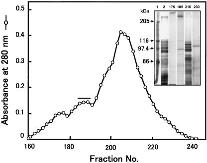
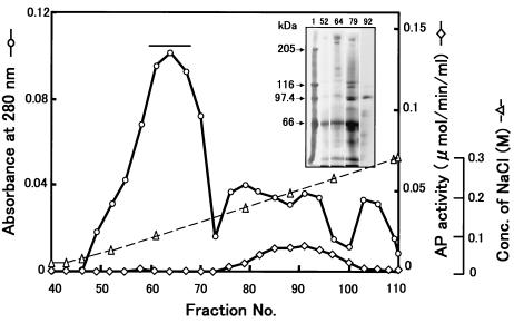
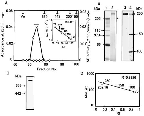
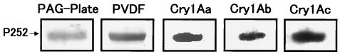
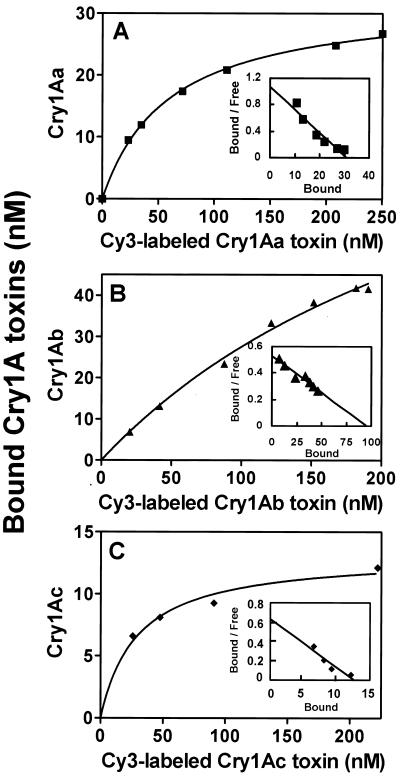
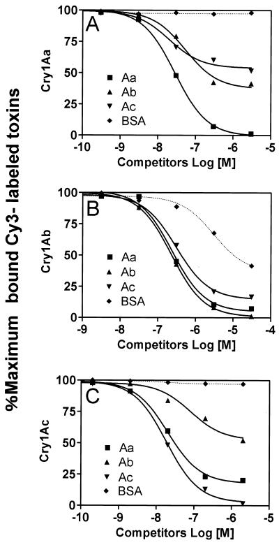
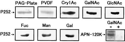
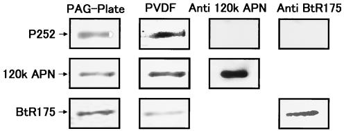
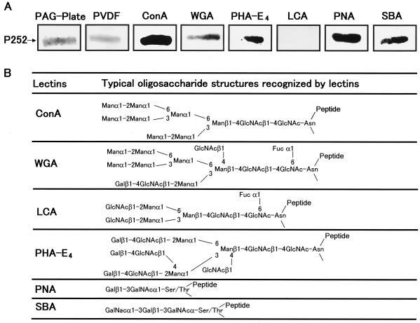
We describe the properties of a novel 252-kDa protein (P252) isolated from brush border membranes of Bombyx mori. P252 was found in a Triton X-100-soluble brush border membrane vesicle fraction, suggesting that it may be a component of the midgut epithelial cell membrane. P252 was purified to homogeneity, and the amino acid sequence of two internal peptides was determined, but neither of the peptides matched protein sequences in the available databases. The apparent molecular mass of the purified protein was estimated by denaturing gel electrophoresis to be 252 kDa, and it migrated as a single band on native gels. However, gel filtration chromatography indicated an apparent mass of 985 kDa, suggesting that P252 may exist as a homo-oligomer. The associations of P252 with Cry1Aa, Cry1Ab, and Cry1Ac were specific, and K(d) constants were determined to be 28.9, 178.5, and 20.0 nM, respectively. A heterologous competition assay was also done. P252 did not exhibit Leu-pNA hydrolysis activity, and binding to the Cry1A toxins was not inhibited by GalNAc. Binding assays of P252 with various lectins indicated the presence of three antennal N-linked high-mannose-type as well as O-linked mucin-type sugar side chains. While the function of P252 is not yet clear, we propose that it may function with Cry1A toxins during the insecticidal response and/or Cry toxin resistance mechanism.


Figures

FIG. 1.
FIG. 1.
Toyopearl HW-65F gel filtration chromatography. Chromatography of BBMV proteins prepared from fifth-instar larvae of B. mori. Each 8 ml of each column fraction was collected at a flow rate of 80 ml/h, and the proteins were resolved by SDS-PAGE (superimposed gel). Lane 1, protein molecular mass markers; lane 2, Triton X-100-soluble protein from BBMV; lane 3, fraction 175; lane 4, fraction 185; lane 5, fraction 210; lane 6, fraction 230. Fractions 185 to 190 (horizontal bar) were pooled and further purified by using DEAE-Sepharose chromatography.
FIG. 2.
FIG. 2.
DEAE-Sepharose chromatography of P252. Chromatogram of DEAE column fractionation of Toyopearl HW-65F gel filtration fractions containing P252. Each 2-ml sample was collected at a flow rate of 60 ml/h from the DEAE column and resolved by SDS-PAGE with silver staining. Lane 1, protein molecular mass markers; lanes 2, fraction 52; lane 3, fraction 64; lane 4, fraction 79; lane 5, fraction 92. Fractions 60 to 68 (horizontal bar) were pooled and further fractionated by DEAE column chromatography and gel filtration chromatography.
FIG. 3.
FIG. 3.
Sephacryl S-300 gel filtration chromatography of P252. (A) Fractions 52 to 64 collected from the DEAE chromatography shown in Fig. 2 were further chromatographed using a Sephacryl S-300 column. The elution profile represents the third of three sequential S-300 fractionation steps. A molecular size calibration standard curve derived using thyroglobulin (669 kDa), apoferritin (443 kDa), β-amylase (200 kDa), and alcohol dehydrogenase (150 kDa) is shown as an inset of panel A. (B) SDS-PAGE of P252 derived from Sephacryl S-300 chromatography from panel A. Lane 1, protein molecular mass markers; lane 2, sample from a pool of fractions 73 to 76 from the absorbance peak designated by the horizontal bar in panel A; lane 3, sample from lane 2 following gel purification; lane 4, protein molecular mass markers. Proteins were visualized by silver staining. (C) Native PAGE of purified P252. The gel was silver stained to determine the purity of the P252 preparation. (D) Determination of molecular weight of P252 by SDS-PAGE. A calibration curve derived from SDS-PAGE molecular weight standards run in parallel with the S-300 fractionated product was used to determine the molecular weight of P252.
FIG. 4.
FIG. 4.
Ligand blot of P252 binding to Cry1A toxins. P252 was resolved by SDS-PAGE and transferred to a PVDF membrane. Samples from the SDS-PAGE gel (PAG-Plate) and the PVDF membrane (PVDF) were stained with CBB. Immobilized P252 was incubated with Cry1Aa, Cry1Ab, and Cry1Ac, and toxin binding was visualized using anti-Cry1Aa and anti-Cry1Ac.
FIG. 5.
FIG. 5.
Total binding assay of Cy3-labeled Cry1A toxins with P252. Binding constants for Cy3-labeled Cry1Aa (A), Cry1Ab (B), and Cry1Ac (C) were determined using a P252 affinity column. Various amounts of Cy3-labeled proteins were incubated on the column and washed. The bound Cry toxin was eluted and quantitated by fluorescence, using a calibration curve. The superimposed graphs show Scatchard plots to determine each kd of Cy3-labeled Cry1A toxin.
FIG. 6.
FIG. 6.
Heterologous competition binding assay between Cry1A toxins and P252. The same procedure was used as for Fig. 5, except that the Cy3-labeled Cry1A toxin solution contained various amounts of competing unlabeled Cry1Aa, Cry1Ab, Cry1Ac, or BSA. The amount of bound Cy3-labeled Cry1A toxin was expressed as a percentage of the maximum labeled Cry1A toxins bound.
FIG. 7.
FIG. 7.
Inhibition of binding between P252 and Cry1Ac by sugars, using ligand blot analysis. P252 was resolved by SDS-PAGE and transferred to a PVDF membrane, and samples from the SDS-PAGE gel (PAG-Plate) and the PDVF membrane (PVDF) were stained with CBB. P252 on the PVDF membrane was preincubated with the sugar GalNAc, GlcNAc, Gal, Fuc, or Man at a 100 mM concentration for an hour and then incubated with Cry1Ac. The sugars used in the blotting assays are shown above each panel. As a positive control, the inhibition of binding between partially purified APN-120K and Cry1Ac was demonstrated with (+) and without (−) GalNAc (bottom-right panel).
FIG. 8.
FIG. 8.
Western blots of P252 using anti-APN-120K and anti-BtR175. P252 was resolved by SDS-PAGE and transferred to a PVDF membrane, and samples from the SDS-PAGE gel (PAG-Plate) and the PDVF membrane (PVDF) were stained with CBB. Western blotting was performed using anti-APN-120K or anti-BtR175, as indicated (top row). As controls, partially purified APN-120K (middle row) and BtR175 (bottom row) were also analyzed using the appropriate antibodies.
FIG. 9.
FIG. 9.
Lectin binding analysis of P252. (A) P252 was resolved by SDS-PAGE and transferred to a PVDF membrane, and samples from the SDS-PAGE gel (PAG-Plate) and the PDVF membrane (PVDF) were stained with CBB. Lectins (ConA, WGA, PHA-E4, LCA, PNA, and SBA) conjugated to peroxidase were incubated with the immobilized P252. (B) Schematic of typical oligosaccharide structures recognized by various lectins used in this experiment.

Similar articles

Cited by

References

    1. Baumann, N. A., and A. K. Menon. 2002. Lipid modification of proteins, p. 37-54. In D. E. Vance and J. E. Vance (ed.), Biochemistry of lipids, lipoproteins and membranes, 4th ed. Elsevier Sciences B. V., Amsterdam, The Netherlands.
    1. Benajiba, A., and S. Maroux. 1981. Subunit structure of pig small-intestinal brush-border aminopeptidase N. Biochem. J. 197:573-580. - PMC - PubMed
    1. Bradford, M. M. 1976. A rapid and sensitive method for the quantitation of microgram quantities of protein utilizing the principle of protein-dye binding. Anal. Biochem. 72:248-254. - PubMed
    1. Cleveland, D. W., S. G. Fischer, M. L. Kirschner, and U. K. Laemmli. 1977. Peptide mapping by limited proteolysis in sodium dodecylsulphate and analysis by gel electrophoresis. J. Biol. Chem. 252:1102-1106. - PubMed
    1. Danielsen, E. M. 1994. Dimeric assembly of enterocyte brush border enzymes. Biochemistry 33:1599-1605. - PubMed

Publication types

MeSH terms

LinkOut - more resources