. 2004 Aug 4;24(31):6904-11.
doi: 10.1523/JNEUROSCI.1978-04.2004.

Synaptic strengthening mediated by bone morphogenetic protein-dependent retrograde signaling in the Drosophila CNS

Affiliations

Synaptic strengthening mediated by bone morphogenetic protein-dependent retrograde signaling in the Drosophila CNS

Richard A Baines. J Neurosci. .

Abstract

Retrograde signaling is an essential component of synaptic development and physiology. Previous studies show that bone morphogenetic protein (BMP)-dependent retrograde signaling is required for the proper development of the neuromuscular junction (NMJ) in Drosophila. These studies, moreover, raised the significant possibility that the development of central motor circuitry might similarly be reliant on such signaling. To test this hypothesis, retrograde signaling between postsynaptic motoneurons and their presynaptic interneurons is examined. Postsynaptic expression of an adenylate cyclase encoded by rutabaga (rut), is sufficient to strengthen synaptic transmission at these identified central synapses. Results are presented to show that the underlying mechanism is dependent on BMP retrograde signaling. Thus, presynaptic expression of an activated TGF-beta receptor, thickvien (tkv), or postsynaptic expression of a TGF-beta ligand, glass-bottom boat (gbb), is sufficient to phenocopy strengthening of synaptic transmission. In the absence of gbb, endogenous synaptic transmission is significantly weakened and, moreover, postsynaptic overexpression of rut is unable to potentiate synaptic function. Potentiation of presynaptic neurotransmitter release, mediated by increased postsynaptic expression of gbb, is dependent on normal cholinergic activity, indicative that either the secretion of this retrograde signal, or its transduction, is activity dependent. Thus, in addition to the development of the NMJ and expression of myoactive FMRFamide-like peptides in specific central neurons, the results of the present study indicate that this retrograde signaling cascade also integrates the development and function of central motor circuitry that controls movement in Drosophila larvae.


Figures
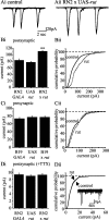
Figure 1.
Figure 1.
Postsynaptic cAMP increases synaptic excitation of aCC/RP2. Ai, Whole-cell voltage-clamp recordings (Vh, -60 mV) from aCC/RP2 reveal large inward currents that are the result of the evoked release of presynaptic ACh (Baines et al., 1999, 2001). Aii, These currents are significantly increased in amplitude after overexpression of rut in aCC/RP2. Each panel shows three successive currents overlayed. Bi, The averaged amplitude of excitatory currents recorded in aCC/RP2 is significantly increased (p ≤ 0.01) after postsynaptic expression of UAS-rut compared with controls (c, parental stocks). Bii, Cumulative probability plots of individual excitatory currents reveals a clear increase in amplitude after overexpression of rut in aCC/RP2. Ci, Cii, Targeted expression of rut to cholinergic neurons (B19-GAL4) does not effect the amplitude of synaptic currents recorded in aCC/RP2. Di, Dii, In the presence of TTX (0.1 μm), evoked excitatory currents are abolished, leaving only those currents that are elicited from the spontaneous release of vesicles in the presynaptic interneurons (Dii, inset; calibration: 4 pA, 0.2 sec). Spontaneous currents in aCC/RP2 are unaffected in amplitude after overexpression of rut in these motoneurons. For all values, n > 8; mean ± SE.
Figure 2.
Figure 2.
Postsynaptic PKA increases synaptic excitation of aCC/RP2. A, Postsynaptic expression (RN2-O GAL4) of a constitutively active PKA transgene (PKAact) increases the amplitude of synaptic excitatory currents recorded in aCC/RP2 (p ≤ 0.01). In contrast, postsynaptic expression of a dominant-negative PKA (PKAinh) reduces the amplitude of these currents (p ≤ 0.01). Values are mean ± SE; n = 8. B, Cumulative probability plots of individual excitatory currents recorded after overexpression of PKAact and PKAinh in aCC/RP2. Control is parental GAL4 and both UAS lines.
Figure 3.
Figure 3.
Presynaptic expression of activated tkv is sufficient to potentiate synaptic transmission. A, Targeted expression (B19-GAL4) of activated tkv (tkvA) is sufficient to markedly increase the amplitude of evoked synaptic currents recorded in aCC/RP2 compared with parental controls (p ≤ 0.001). Parental UAS-tkvA stocks also exhibit significantly increased synaptic currents compared with B19-GAL4 (p ≤ 0.01). By comparison, GAL4-mediated expression of activated sax (saxA) shows no further increase in synaptic current amplitude above that seen in UAS-saxA controls (which again are significantly increased compared with B19-GAL4; p ≤ 0.01). B, Cumulative probability plots show a marked shift to larger amplitudes for almost all currents recorded after GAL4-mediated expression of tkvA compared with parental controls (GAL4/UAS). A similar plot for spontaneous mEPSC amplitudes shows no such significant differences (inset). C, Postsynaptic expression (RN2-GAL4) of either tkvA or saxA does not increase synaptic current amplitude in aCC/RP2, above that seen in UAS controls. For all values, n > 8; mean ± SE.
Figure 4.
Figure 4.
Postsynaptic expression of gbb is sufficient to increase the amplitude of synaptic currents. A, Targeted expression (RN2-GAL4) of gbb in aCC/RP2 is sufficient to greatly increase the amplitude of evoked synaptic currents recorded (p ≤ 0.001), compared with the UAS control, which is also significantly increased compared with RN2-GAL4 (p ≤ 0.01). B, Cumulative probability plots show a large shift to the right for evoked currents observed in aCC/RP2 that are overexpressing gbb compared with control (GAL4/UAS). In the presence of TTX, spontaneous mEPSC amplitudes are, in contrast, significantly smaller because of overexpression of gbb in aCC/RP2 (inset). C, Targeted expression of gbb to cholinergic neurons (B19-GAL4) fails to increase synaptic current amplitude in aCC/RP2, above that already observed for UAS-gbb alone. For all values, n > 8; mean ± SE.
Figure 5.
Figure 5.
Gbb is necessary for synaptic strengthening. A, Evoked synaptic current amplitude recorded in aCC/RP2 is significantly smaller in null alleles of gbb (gbb1 and gbb2) compared with heterozygous controls (allele/Cyo::GFP balancer; p ≤ 0.01). Moreover, postsynaptic expression of rut, which is normally sufficient to significantly increase evoked synaptic currents in aCC/RP2 (Fig. 1 A,B), fails to do so in the absence of gbb. B, Cumulative plots of evoked synaptic current amplitude reinforce the observation that in the absence of gbb, targeted expression (RN2-GAL4) of rut in aCC/RP2 fails to increase current amplitude above that seen in either null allele of gbb examined (gbb1 shown).
Figure 6.
Figure 6.
Gbb signaling is activity dependent. A, After a 3 hr period of egg laying at 20°C, chats2 embryos were shifted to 29°C and allowed to develop to late stage 17 (Baines et al., 2001). Before recording synaptic currents in aCC/RP2, embryos were transferred back to 20°C for an additional 2 hr, during which time hatching occurred. Recordings of current amplitude under these conditions reveal that postsynaptic expression of gbb (using RN2-O GAL4) is unable to potentiate synaptic current amplitude in the absence of cholinergic signaling. By comparison, heterozygous controls (chats2/FM7), in which cholinergic signaling is functional, which also carried both RN2-O GAL4 and UAS-gbb, showed significantly larger synaptic currents compared with chats2 (p ≤ 0.01). B, Restoration of cholinergic function for extended periods (∼22 hr) in these same larvae is sufficient to restore the ability of postsynaptic expression of gbb to potentiate synaptic current amplitude (identical letters indicate p ≤ 0.01). For all values, n > 6; mean ± SE.

Similar articles

Cited by

References

    1. Aberle H, Haghighi AP, Fetter RD, McCabe BD, Magalhães TR, Goodman CS (2002) Wishful thinking encodes a BMP type II receptor that regulates synaptic growth in Drosophila. Neuron 33: 545-558. - PubMed
    1. Allan DW, St Pierre SE, Miguel-Aliaga I, Thor S (2003) Specification of neuropeptide cell identity by the integration of retrograde BMP signalling and a combinatorial transcription factor code. Cell 113: 73-86. - PubMed
    1. Baines RA (2003) Postsynaptic protein kinase A reduces neuronal excitability in response to increased synaptic excitation in the Drosophila CNS. J Neurosci 23: 8664-8672. - PMC - PubMed
    1. Baines RA, Bate M (1998) Electrophysiological development of central neurons in the Drosophila embryo. J Neurosci 18: 4673-4683. - PMC - PubMed
    1. Baines RA, Robinson SG, Fujioka M, Jaynes JB, Bate M (1999) Postsynaptic vesicle release is essential for synaptogenesis in Drosophila. Curr Biol 9: 1267-1270. - PMC - PubMed

Publication types

LinkOut - more resources