. 2004 Aug 4;24(31):6920-7.
doi: 10.1523/JNEUROSCI.0473-04.2004.

Glutamate released from glial cells synchronizes neuronal activity in the hippocampus

Affiliations

Glutamate released from glial cells synchronizes neuronal activity in the hippocampus

María Cecilia Angulo et al. J Neurosci. .

Erratum in

  • J Neurosci. 2004 Aug 25;24(34):2p following 7575

Abstract

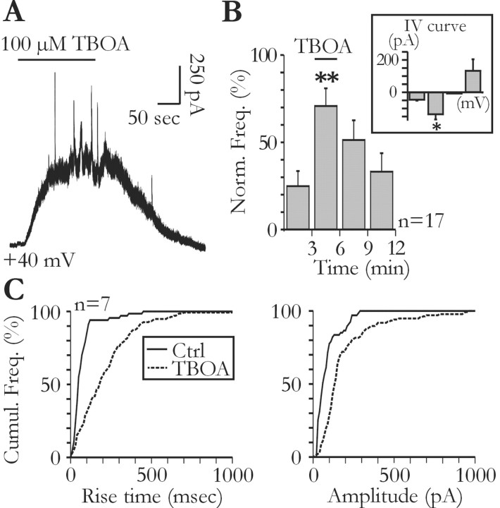
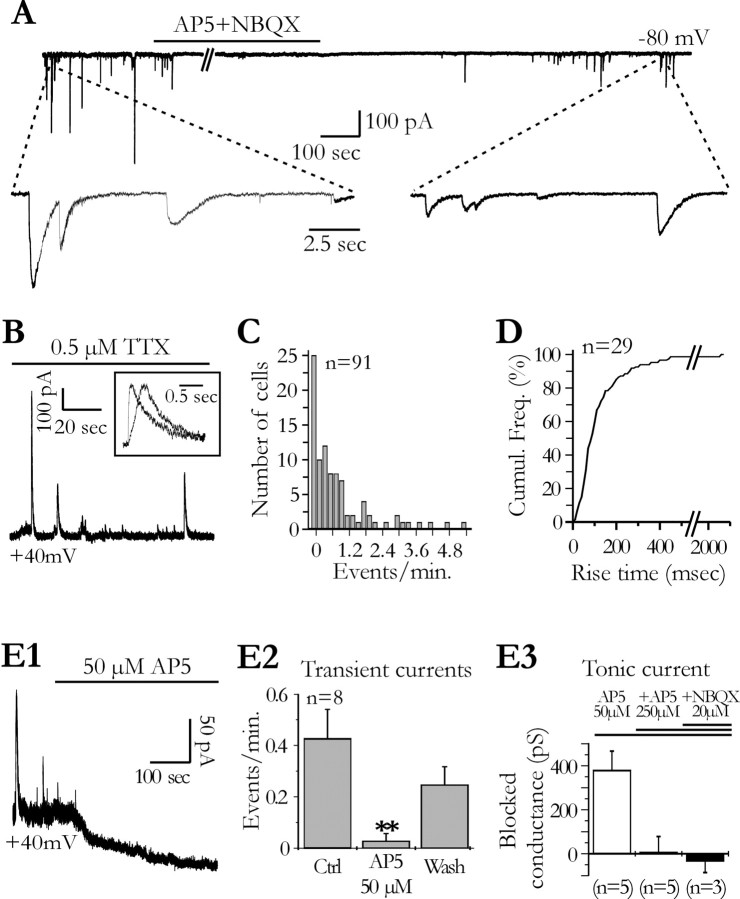
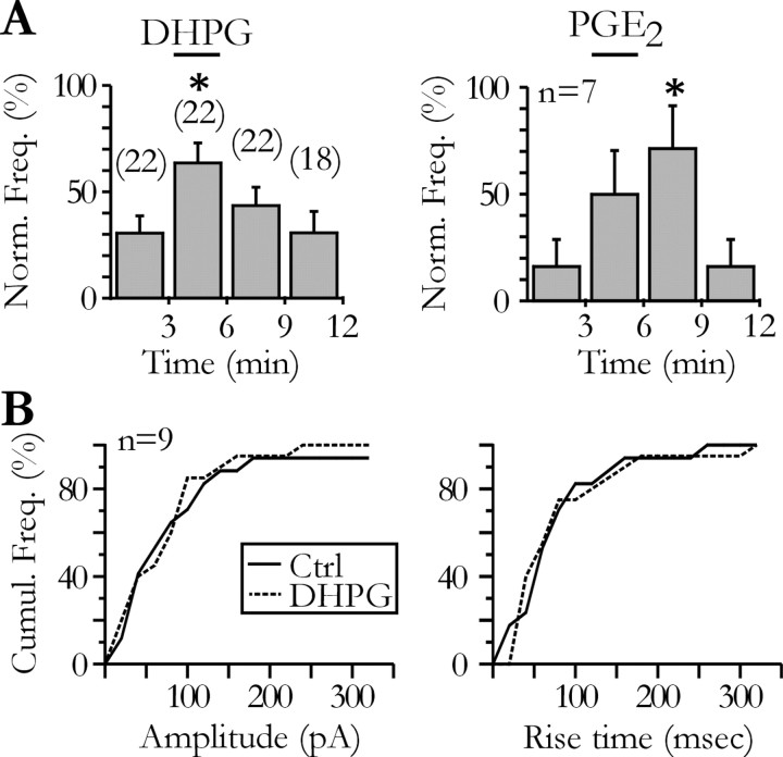
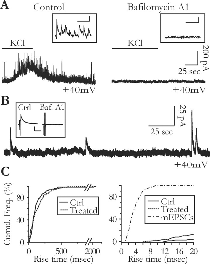
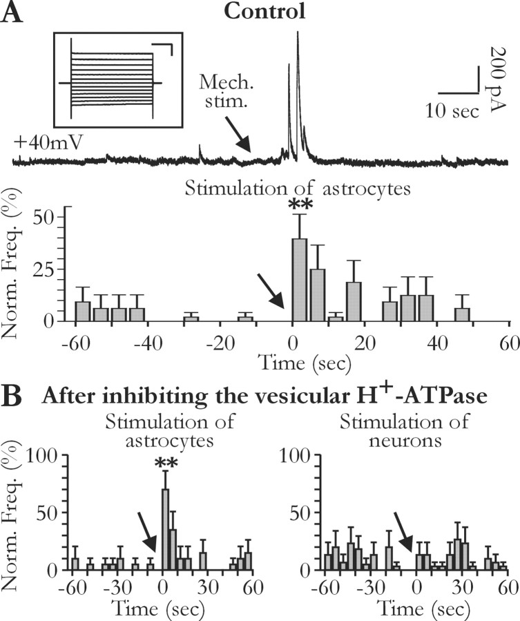
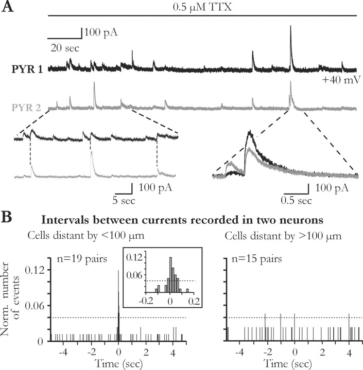
Glial cells of the nervous system directly influence neuronal and synaptic activities by releasing transmitters. However, the physiological consequences of this glial transmitter release on brain information processing remain poorly understood. We demonstrate here in hippocampal slices of 2- to 5-week-old rats that glutamate released from glial cells generates slow transient currents (STCs) mediated by the activation of NMDA receptors in pyramidal cells. STCs persist in the absence of neuronal and synaptic activity, indicating a nonsynaptic origin of the source of glutamate. Indeed, STCs occur spontaneously but can also be induced by pharmacological tools known to activate astrocytes and by the selective mechanical stimulation of single nearby glial cells. Bath application of the inhibitor of the glutamate uptake dl-threo-beta-benzyloxyaspartate increases both the frequency of STCs and the amplitude of a tonic conductance mediated by NMDA receptors and probably also originated from glial glutamate release. By using dual recordings, we observed synchronized STCs in pyramidal cells having their soma distant by <100 microm. The degree of precision (<100 msec) of this synchronization rules out the involvement of calcium waves spreading through the glial network. It also indicates that single glial cells release glutamate onto adjacent neuronal processes, thereby controlling simultaneously the excitability of several neighboring pyramidal cells. In conclusion, our results show that the glial glutamate release occurs spontaneously and synchronizes the neuronal activity in the hippocampus.


Figures

Figure 5.
Figure 5.
Glial glutamate release contributes to ambient glutamate concentration. A, Tonic and slow transient currents are unmasked by bath application of TBOA (100 μm). B, Effect of TBOA on the frequency of slow transient currents (ANOVA test; p < 0.001). The voltage dependency of slow transient currents during the application of TBOA was obtained with 1 mm Ca2+ and 1 mm Mg2+ in the extracellular solution (inset). The bars from left to right correspond to the mean amplitude of transient currents measured at -70, -30, 0, and +30 mV (n = 3; Student's t test, p < 0.05). C, The cumulative distributions of the rise time (left) and amplitude (right) of the slow transient currents are shifted to the right during TBOA application (Kolmogorov-Smirnov test; p < 0.001). Ctrl, Control.
Figure 1.
Figure 1.
Slow transient and tonic NMDA receptor-mediated currents in hippocampal pyramidal cells. A, Spontaneous slow transient currents recorded at a holding potential of -80 mV are blocked by d-AP-5 (300 μm) and NBQX (20 μm). The frequency of the slow transient currents in this cell (2.5 events/min) before the application of the antagonists is in the top range of the values measured in our sample. B, In another neuron, spontaneous slow transient currents are recorded at +40 mV in the presence of TTX. Two currents normalized in amplitude are shown in the inset to illustrate the variability of the kinetics. C, Distribution of the transient current mean frequency for 91 neurons recorded at +40 mV in the presence of TTX (0.5-1 μm). Note the low frequency of these currents. D, Cumulative frequency distribution of the rise time (t10-90%) of the slow transient currents for 29 neurons held at +40 mV in the presence of TTX. E, Bath application of d-AP-5 abolishes slow transient currents and a tonic current. E1, Effects of 50 μm d-AP-5 on the membrane current of a CA1 pyramidal cell recorded at +40 mV. E2, Effect of d-AP-5 on the frequency of the slow transient currents (ANOVA test; p < 0.01). E3, Mean tonic conductance blocked by 50 μm d-AP-5 and the absence of effect of a higher concentration of d-AP-5 and of NBQX. Ctrl, Control; Wash, recovery after washing out d-AP-5.
Figure 2.
Figure 2.
DHPG and PGE2 increase the frequency of the slow transient currents recorded in pyramidal neurons. A, Effect of bath applications of DHPG (10 μm; left) and PGE2 (5-20 μm; right) on the frequency of the slow transient currents in neurons held at -80 mV (ANOVA test; p < 0.05). Numbers in parenthesis above the bars of the left histogram refer to the number of tested pyramidal neurons. B, Comparison of the cumulative frequency distributions of the amplitude (left) and of the 10-90% rise time (right) of the STCs recorded before (Ctrl) and during the application of DHPG (10 μm) for nine neurons showing both spontaneous and agonist-evoked STCs (Kolmogorov-Smirnov tests; p > 0.05). Ctrl, Control.
Figure 3.
Figure 3.
Slow transient currents persist in the absence of neuronal vesicular release. A, Preincubation of the slice with the vacuolar H+-ATPases inhibitor bafilomycin A1 (4 μm; right) inhibits the frequency increase of synaptic currents induced by 10 mm extracellular potassium normally observed in control slices (left). The insets show a portion of the traces during the increase in potassium concentration at a faster time scale. Calibration: 100 msec, 50 pA. B, Preincubation of the slices with 4 μm bafilomycin A1 does not inhibit the slow transient currents. The inset shows that incubation of the slices with bafilomycin A1 (Baf. A1) abolishes the synaptic responses, which can be evoked by the electrical stimulation of Schaffer collaterals in nontreated slices (Ctrl). The stimulation intensity for the control and the pretreated slice is 0.4 and 1 mA, respectively. Calibration: 500 msec, 100 pA. C, Preincubation of the slices with 4 μm bafilomycin A1 or 2 μm concanamycin A does not change the kinetics of the slow transient currents (left: Ctrl, same control plot as in Fig. 1 D; Treated, 12 pretreated slices, n = 12 cells; Kolmogorov-Smirnov test; p > 0.05; right: mEPSC distribution obtained from 3 neurons recorded in nontreated slices and superimposed with the beginning of the STC distributions shown on the right panel).
Figure 4.
Figure 4.
Mechanical stimulations of glial cells induce slow transient currents in hippocampal pyramidal neurons. A, The top trace illustrates slow transient currents induced by the mechanical stimulation (arrow, Mech. stim.) with a patch pipette of a single glial cell located close to the recorded neuron (<50 μm). The inset illustrates a voltage-clamp recording in response to pulses from -150 to -30 mV of the glial cell stimulated mechanically during the recording of this neuron (resting membrane potential, -91 mV). Calibration: 100 msec, 0.5 nA. The histogram shows the grouped data for 11 neuronal recordings, during which a total of 23 glial cells was stimulated. The frequency of the transient currents increased significantly during the 5 sec after glial cell stimulations (ANOVA test; p < 0.001). B, Effect of mechanical stimulations of single glial cells (left) and neurons (right) after preincubation of the slices with 4 μm bafilomycin A1 or 2 μm concanamycin A. Note that the frequency of the transient currents does not increase when neurons are mechanically stimulated (left: 12 stimulated glial cells and 4 recorded neurons; right: 11 stimulated neurons and 7 recorded neurons; ANOVA test, p < 0.001 and p > 0.05, respectively).
Figure 6.
Figure 6.
Glutamate released from glial cells synchronizes neuronal activity. A, Slow transient currents are synchronized in two different pyramidal cells recorded simultaneously in the presence of TTX. Note the high degree of precision of the synchronization. B, Normalized distributions of current time intervals in neuronal pairs for which the somata were distant by less (left) or more (right) than 100 μm. The noise level is indicated by the dashed line. Note that synchronized events occur only when neurons are distant by <100 μm (inset). Number of events: left, 84; right, 43.

Similar articles

Cited by

References

    1. Aguado F, Espinosa-Parrilla JF, Carmona MA, Soriano E (2002) Neuronal activity regulates correlated network properties of spontaneous calcium transients in astrocytes in situ J Neurosci 22: 9430-9444. - PMC - PubMed
    1. Araque A, Parpura V, Sanzgiri RP, Haydon PG (1998a) Glutamate-dependent astrocyte modulation of synaptic transmission between cultured hippocampal neurons. Eur J Neurosci 10: 2129-2142. - PubMed
    1. Araque A, Sanzgiri RP, Parpura V, Haydon PG (1998b) Calcium elevation in astrocytes causes an NMDA receptor-dependent increase in the frequency of miniature synaptic currents in cultured hippocampal neurons. J Neurosci 18: 6822-6829. - PMC - PubMed
    1. Araque A, Parpura V, Sanzgiri RP, Haydon PG (1999) Tripartite synapses: glia, the unacknowledged partner. Trends Neurosci 22: 208-215. - PubMed
    1. Araque A, Li N, Doyle RT, Haydon PG (2000) SNARE protein-dependent glutamate release from astrocytes. J Neurosci 20: 666-673. - PMC - PubMed

Publication types

Substances

LinkOut - more resources