. 2004 Aug;16(8):2204-16.
doi: 10.1105/tpc.104.021790.

Tissue-specific expression of head-to-tail cyclized miniproteins in Violaceae and structure determination of the root cyclotide Viola hederacea root cyclotide1

Affiliations

Tissue-specific expression of head-to-tail cyclized miniproteins in Violaceae and structure determination of the root cyclotide Viola hederacea root cyclotide1

Manuela Trabi et al. Plant Cell. 2004 Aug.

Abstract

The plant cyclotides are a family of 28 to 37 amino acid miniproteins characterized by their head-to-tail cyclized peptide backbone and six absolutely conserved Cys residues arranged in a cystine knot motif: two disulfide bonds and the connecting backbone segments form a loop that is penetrated by the third disulfide bond. This knotted disulfide arrangement, together with the cyclic peptide backbone, renders the cyclotides extremely stable against enzymatic digest as well as thermal degradation, making them interesting targets for both pharmaceutical and agrochemical applications. We have examined the expression patterns of these fascinating peptides in various Viola species (Violaceae). All tissue types examined contained complex mixtures of cyclotides, with individual profiles differing significantly. We provide evidence for at least 57 novel cyclotides present in a single Viola species (Viola hederacea). Furthermore, we have isolated one cyclotide expressed only in underground parts of V. hederacea and characterized its primary and three-dimensional structure. We propose that cyclotides constitute a new family of plant defense peptides, which might constitute an even larger and, in their biological function, more diverse family than the well-known plant defensins.


Figures

Figure 1.
Figure 1.
Amino Acid Sequences of Various Cyclotides Representative of the Bracelet and Möbius Subfamilies. The amino acids are colored according to their properties (hydrophobic, pink; hydrophilic, green; acidic, red; basic, blue). Cys residues are given in yellow, and intercysteine loops are numbered at top of figure. The yellow lines on the bottom of the figure indicate the disulfide connectivities. The subfamilies can be distinguished by the presence (Möbius) or absence (bracelet) of a cis-Pro residue in loop 5. Note also the loop sizes (loop 3) and loop sequences characteristic for each subfamily. As a result of the cyclic nature of the peptide backbone in cyclotides and some ambiguity about precursor processing sites and mechanism(s), the numbering of residues is arbitrary, starting with the first absolutely conserved residue in the mature cyclotide sequence (Cys1) in the loop with highest conservation within and across the two subfamilies. The sequences have been aligned using MultAlin (Corpet, 1988) with Blosum62, a gap opening penalty of 10 and a gap extension penalty of 2, followed by a manual alignment of the structurally crucial hydroxyl bearing residue in loop 3 (Rosengren et al., 2003) The amino acid sequence and residue numbering of vhr1, determined in this study, are given at bottom of figure. Original citations to the various cyclotides are as follows: kalata B1, Saether et al. (1995); kalata B2, kalata B5, cycloviolacin O1, and cycloviolacin O10, Craik et al. (1999); kalata B6, Jennings et al. (2001); varv F, Göransson et al. (1999); cyclopsychotride A, Witherup et al. (1994); circulin A and circulin B, Gustafson et al. (1994); circulin E and circulin F, Gustafson et al. (2000); cycloviolin B, Hallock et al. (2000); and vhr1, this study. A complete list of cyclotide sequences and database access IDs is given as supplemental material (see Supplemental Table S1 online).
Figure 2.
Figure 2.
Effects of Disulfide Bond Modification on the LC-MS Profile of Cyclotides. LC-MS profile of a native cyclotide fraction (A). After reduction and alkylation of the disulfide bonds (B), the modified cyclotides elute significantly earlier and show masses that correspond to six alkylated half-cystines, providing proof that three disulfide bonds have been broken. (C) provides a tabular listing of the masses observed in (A) (column native) together with the calculated [red/alk (calc)] and observed [red/alk (obs)] masses of the reduced and alkylated peptides. Note that reduction and alkylation of such a complex reaction mixture can lead to incomplete reaction of some species; therefore, masses corresponding to some native cyclotides found in (A) are missing in (B) and (C).
Figure 3.
Figure 3.
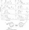
LC-MS Profiles of Cyclotides in Various V. hederacea Plant Parts and Sequencing Strategy for vhr1. (A) to (H) LC-MS profiles of various parts of V. hederacea. Leaves (A); petioles (B); flowers (C); pedicels (D); aboveground runners (E); bulbs (F); belowground runners (G); and roots (H). The peaks are labeled with the masses of the cyclotides they contain. The presence of one or more minor components with a signal intensity of <30% of that of the strongest corresponding signal is indicated by a plus symbol (+); the masses of these components are not given in the figure. For peaks that are identified by a symbol, the masses are given elsewhere in the figure panel, preceded by the corresponding symbol. The peaks containing kalata B1 (kb1) and vhr1 are labeled ([C] and [H], respectively). (I) Characterization of vhr1. The disulfide bonds of the native, circular peptide are reduced and alkylated to destabilize the fold. Enzymatic digest with endoproteinase GluC yields a linear peptide whose amino acid sequence can be determined by Edman degradation.
Figure 4.
Figure 4.
LC-MS Profiles of Aerial and Underground Parts of Various Viola Species. For labeling of the peaks, see Figure 3. The aerial parts of V. odorata contain a further cyclotide peak not shown in the profile (elution time 22.3 min, mass of the main compound 2748 D).
Figure 5.
Figure 5.
Structural Characteristics of vhr1. (A) Structural family of vhr1. Stereo view of the 20 lowest energy structures of vhr1 superimposed over the N, C, and Cα atoms of all 30 residues. The peptide backbone is given in blue, and the disulfide bonds are in yellow. (B) Summary of the structural features present in vhr1. The disulfide bonds are given in yellow, and the intercysteine loops are labeled. (C) The cystine knot motif in vhr1. The atoms of the cystine knot are scaled according to their van der Waals radii. The two disulfide bonds and the backbone segments forming the ring are shown in blue and yellow, respectively. The third disulfide bond, penetrating this disulfide/backbone ring, is shown in orange. Backbone segments not directly involved in the cystine knot motif are shown as lines with labeled loops.
Figure 6.
Figure 6.
Surface Representation of the Lowest Energy Structure of vhr1 in Comparison with Kalata B1. The amino acids are colored according to their properties (red, acidic; blue, basic; green, hydrophilic; gray, hydrophobic). Cys residues are given in yellow, and Gly residues are in light gray. Selected residues are labeled with the one letter code and the residue number, following the numbering of residues in the respective PDB entries.

Similar articles

Cited by

References

    1. Barry, D.G., Daly, N.L., Bokesch, H.R., Gustafson, K.R., and Craik, D.J. (2004). Solution structure of the cyclotide palicourein. Implications for the development of a pharmaceutical framework. Structure 12, 85–94. - PubMed
    1. Bax, A., and Davies, D.G. (1985). MLEV-17 based two dimensional homonuclear magnetisation transfer spectroscopy. J. Magn. Reson. 65, 355–360.
    1. Bode, W., Greyling, H.J., Huber, R., Otlewski, J., and Wilusz, T. (1989). The refined 2.0 A X-ray crystal structure of the complex formed between bovine beta-trypsin and CMTI-I, a trypsin inhibitor from squash seeds (Cucurbita maxima). Topological similarity of the squash seed inhibitors with the carboxypeptidase A inhibitor from potatoes. FEBS Lett. 242, 285–292. - PubMed
    1. Bokesch, H.R., Pannell, L.K., Cochran, P.K., Sowder, R.C.I., McKee, T.C., and Boyd, M.R. (2001). A novel anti-HIV macrocyclic peptide from Palicourea condensata. J. Nat. Prod. 64, 249–250. - PubMed
    1. Boman, H.G. (1995). Peptide antibiotics and their role in innate immunity. Annu. Rev. Immunol. 13, 61–92. - PubMed

Publication types

Associated data

LinkOut - more resources