. 2004 Sep;13(9):2330-40.
doi: 10.1110/ps.04787204. Epub 2004 Aug 4.

Arginine 60 in the ArsC arsenate reductase of E. coli plasmid R773 determines the chemical nature of the bound As(III) product

Affiliations

Arginine 60 in the ArsC arsenate reductase of E. coli plasmid R773 determines the chemical nature of the bound As(III) product

Srini DeMel et al. Protein Sci. 2004 Sep.

Abstract

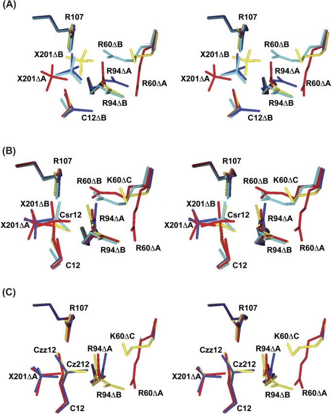
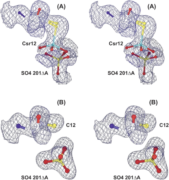
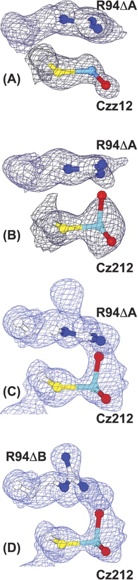
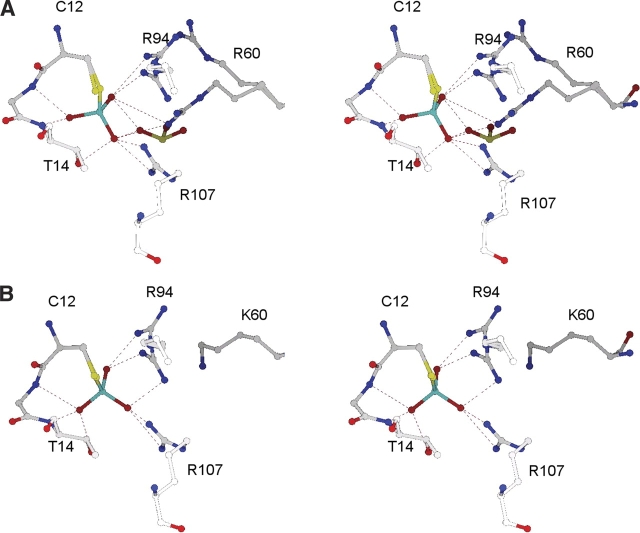
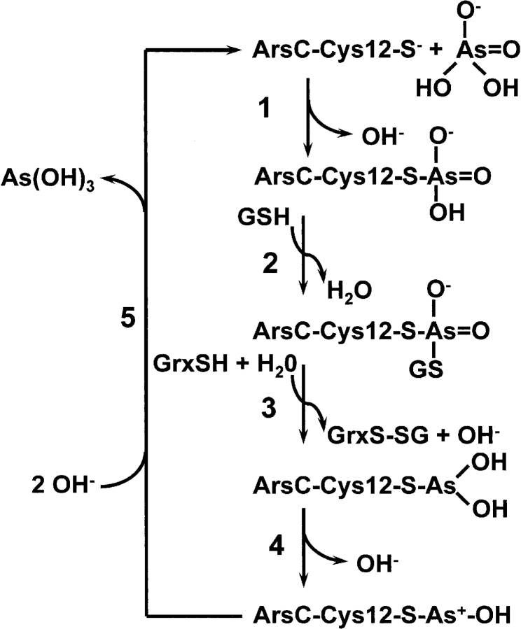
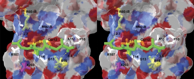
Arsenic is a ubiquitous environmental toxic metal. Consequently, organisms detoxify arsenate by reduction to arsenite, which is then excreted or sequestered. The ArsC arsenate reductase from Escherichia coli plasmid R773, the best characterized arsenic-modifying enzyme, has a catalytic cysteine, Cys 12, in the active site, surrounded by an arginine triad composed of Arg 60, Arg 94, and Arg 107. During the reaction cycle, the native enzyme forms a unique monohydroxyl Cys 12-thiol-arsenite adduct that contains a positive charge on the arsenic. We hypothesized previously that this unstable intermediate allows for rapid dissociation of the product arsenite. In this study, the role of Arg 60 in product formation was evaluated by mutagenesis. A total of eight new structures of ArsC were determined at resolutions between 1.3 A and 1.8 A, with R(free) values between 0.18 and 0.25. The crystal structures of R60K and R60A ArsC equilibrated with the product arsenite revealed a covalently bound Cys 12-thiol-dihydroxyarsenite without a charge on the arsenic atom. We propose that this intermediate is more stable than the monohydroxyarsenite intermediate of the native enzyme, resulting in slow release of product and, consequently, loss of activity.


Figures

Figure 1.
Figure 1.
Ligands in the active site. The active site around Cys 12 is shown in divergent stereo for the native (red), C12S (cyan), R60A (blue), and R60K (yellow) structures in 3.1 M sulfate ion and (A) no other ligand, (B) 0.4 M sodium arsenate, and (C) 0.4 M sodium arsenite. X201 is SO4201.
Figure 2.
Figure 2.
Electron density for arsenate in the active site. (A) Arsenate forms a covalent adduct (Csr12) with cysteine 12 in a fraction of the R60K ArsC molecules in the crystal (structure V). The oxyanion, SO4201Δ, is present only in the fraction of the enzyme molecules that has no adduct. (B) The oxyanion (sulfate or arsenate) binds noncovalently to R60A ArsC (structure VIII). The 2FoFc electron density, shown in divergent stereo, is contoured at 1.0 σ.
Figure 3.
Figure 3.
Electron density for arsenite in the active site. The 2Fo − Fc electron density for the thiarsadihydroxy adducts in crystals of (A) native ArsC (PDB code 1I9D), (B) R60K ArsC, (C) R60A ArsC with Arg94ΔA, and (D) R60A with Arg94ΔB. The electron density was contoured at 1.0 σ.
Figure 4.
Figure 4.
Interactions of the Csr12 arsonocysteine adducts. Csr12 and its adjacent residues within 4.0 Å are shown for (A) native ArsC and (B) the R60K mutant. The hydrogen bonds are depicted with dotted lines. The cyan atom in Csr12 is arsenic.
Figure 5.
Figure 5.
Reaction mechanism of the R733 ArsC arsenate reductase. The mechanism is consistent with the crystal structures described in Table 1. In step 1, the free enzyme (structure I) forms the observed covalent intermediate with arsenate (Martin et al. 2001). In step 2, this intermediate is glutathionylated, a structure that has not yet been obtained. In step 3, As(V) is reduced to As(III), producing a dihydroxy arsenite intermediate (structures VI, IX). In step 4, the novel monohydroxy intermediate with a positively charged arsenic is formed (Martin et al. 2001). Finally, in step 5, the free enzyme is regenerated (structure I).
Figure 6.
Figure 6.
Model of glutathione covalently bound to Csr12. GSH (modified from PDB code 1LBK), is depicted in divergent stereo (green tubes) attached to Csr12 in place of O3. The two carboxyl ends of GSH can form hydrogen bonds (yellow arrows) with Arg60ΔB (2.7 Å) and with Arg 16 (3.3 Å). Csr12, Arg60ΔB, and Arg 16 are colored yellow. Other residues within 4.0 Å of GSH are colored white. The transparent surface delineates the van der Waals surface of ArsC. Although GSH has no steric conflicts with any ArsC residues, it does clash with SO4201 and SO4202, which are omitted. SO4203 is shown in pink (X203).

Similar articles

Cited by

References

    1. Bennett, M.S., Guan, Z., Laurberg, M., and Su, X.D. 2001. Bacillus subtilis arsenate reductase is structurally and functionally similar to low molecular weight protein tyrosine phosphatases. Proc. Natl. Acad. Sci. 98 13577–13582. - PMC - PubMed
    1. Carugo, O. and Bordo, D. 1999. How many water molecules can be detected by protein crystallography? Acta Crystallogr. D Biol. Crystallogr. 55 479–483. - PubMed
    1. Chen, C.M., Misra, T.K., Silver, S., and Rosen, B.P. 1986. Nucleotide sequence of the structural genes for an anion pump. The plasmid-encoded arsenical resistance operon. J. Biol. Chem. 261 15030–15038. - PubMed
    1. Fersht, A. 1999. Structure and mechanism in protein science: A guide to enzyme catalysis and protein folding. W.H. Freeman, New York.
    1. Gladysheva, T.B., Oden, K.L., and Rosen, B.P. 1994. Properties of the arsenate reductase of plasmid R773. Biochemistry 33 7288–7293. - PubMed

Publication types

MeSH terms

LinkOut - more resources