. 2004 Aug;42(8):3406-14.
doi: 10.1128/JCM.42.8.3406-3414.2004.

High genetic diversity of the attachment (G) protein of human metapneumovirus

Affiliations

High genetic diversity of the attachment (G) protein of human metapneumovirus

Nobuhisa Ishiguro et al. J Clin Microbiol. 2004 Aug.

Abstract

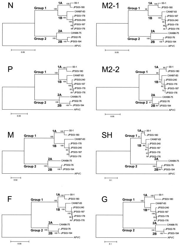
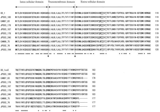
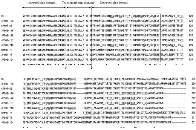
Complete genes encoding the predicted nucleoprotein (N), phosphoprotein (P), matrix protein (M), fusion protein (F), M2-1protein, M2-2protein, small hydrophobic protein (SH), and attachmentprotein (G) of seven newly isolated human metapneumoviruses (hMPVs) were analyzed and compared with previously published data for hMPV genes. Phylogenetic analysis of the nucleotide sequences indicated that there were two genetic groups, tentatively named groups 1 and 2, similar to the grouping of human respiratory syncytial virus. Although the predicted amino acid sequences of N, P, M, F, and M2 were highly conserved between the two groups (amino acid identities, 96% for N, 85% for P, 97% for M, 94% for F, 95% for M2-1, and 90% for M2-2), the amino acid identities of the SH and G proteins were low (SH, 58%; G, 33%). Furthermore, each group could be subdivided into two subgroups by phylogenetic analysis, tentatively named subgroups 1A and 1B and subgroups 2A and 2B. The predicted amino acid sequences of G within members of each subgroup were highly conserved (amino acid identities, 88% for group 1A, 93% for group 1B, and 96% for group 2B). The G of hMPV is thought to be the major antigenic determinant and to play an important role in the production of neutralizing antibodies. Clarification of the antigenic diversity of G is important for epidemiological analysis and for establishment of strategies to prevent hMPV infection.


Figures

FIG. 1.
FIG. 1.
Phylogenetic analysis of hMPV isolates. The putative ORFs for N, P, M, F, M2-1, M2-2, SH, and G were analyzed. The corresponding gene sequences from APVC were also analyzed. Bootstrap proportions were plotted at the main internal branches of the phylogram to show support values. Note that the scale of each tree was different. Phylogenetic analysis was performed by use of the neighbor-joining method of the MEGA program.
FIG. 2.
FIG. 2.
Comparison of the predicted amino acid sequences of SH of hMPV isolates. The predicted amino acid sequence of SH of hMPV with cysteine residues is shown in boldface type, potential N-linked glycosylation sites are shaded in gray, and potential O-glycosylation sites are underlined. Dashes indicate gaps introduced to maximize the alignment or to denote the absence of corresponding amino acids. The asterisks underneath each alignment denote amino acid identity among all sequences. Proposed intracellular, transmembrane, and extracellular domains are indicated above the sequences.
FIG. 3.
FIG. 3.
Comparison of the predicted amino acid sequences of G of hMPV isolates. The predicted amino acid sequence of G of hMPV with cysteine residues is shown in boldface type, potential N-linked glycosylation sites are shaded in gray, and potential O-glycosylation sites are underlined. Dashes indicate gaps introduced to maximize the alignment or to denote the absence of corresponding amino acids. The asterisks underneath each alignment denote amino acid identity among all sequences. Proposed intracellular, transmembrane, and extracellular domains are indicated above the sequences. The square indicates the positions of conserved amino acid residues.
FIG. 4.
FIG. 4.
Predicted gene boundaries, cis-acting gene-start and gene-end signals, and intergenic regions of hMPV isolates. The alignments show the N-P, P-M, M-F, F-M2, M2-SH, SH-G, and G boundaries. Conserved sequence motifs at the end and beginning of each gene are indicated by bold uppercase letters, and a consensus sequence is given below. Translational stop and start codons are underlined.

References

    1. Altschul, S. F., T. L. Madden, A. A. Schaffer, J. Zhang, Z. Zhang, W. Miller, and D. J. Lipman. 1997. Gapped BLAST and PSI-BLAST: a new generation of protein database search programs. Nucleic Acids Res. 25:3389-3402. - PMC - PubMed
    1. Bastien, N., S. Normand, T. Taylor, D. Ward, T. C. Peret, G. Boivin, L. J. Anderson, and Y. Li. 2003. Sequence analysis of the N, P, M and F genes of Canadian human metapneumovirus strains. Virus Res. 93:51-62. - PMC - PubMed
    1. Bastien, N., D. Ward, P. Van Caeseele, K. Brandt, S. H. Lee, G. McNabb, B. Klisko, E. Chan, and Y. Li. 2003. Human metapneumovirus infection in the Canadian population. J. Clin. Microbiol. 41:4642-4646. - PMC - PubMed
    1. Biacchesi, S., M. H. Skiadopoulos, G. Boivin, C. T. Hanson, B. R. Murphy, P. L. Collins, and U. J. Buchholz. 2003. Genetic diversity between human metapneumovirus subgroups. Virology 315:1-9. - PubMed
    1. Boivin, G., Y. Abed, G. Pelletier, L. Ruel, D. Moisan, S. Cote, T. C. Peret, D. D. Erdman, and L. J. Anderson. 2002. Virological features and clinical manifestations associated with human metapneumovirus: a new paramyxovirus responsible for acute respiratory-tract infections in all age groups. J. Infect. Dis. 186:1330-1334. - PubMed

Publication types

LinkOut - more resources