. 2004 Aug 17;101(33):12207-11.
doi: 10.1073/pnas.0404650101. Epub 2004 Aug 5.

Autocatalytic self-propagation of misfolded prion protein

Affiliations

Autocatalytic self-propagation of misfolded prion protein

Jan Bieschke et al. Proc Natl Acad Sci U S A. .

Abstract

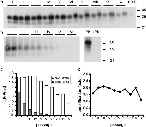
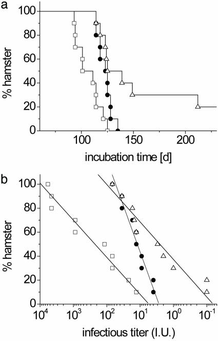
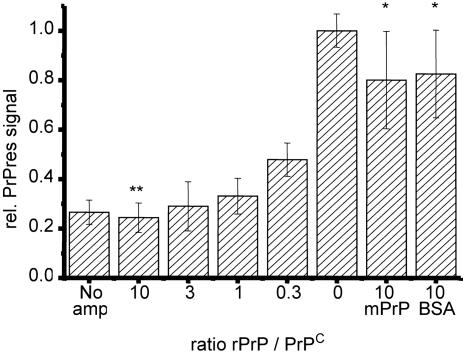
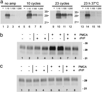
Prions are thought to replicate in an autocatalytic process that converts cellular prion protein (PrP(C)) to the disease-associated misfolded PrP isoform (PrP(Sc)). Our study scrutinizes this hypothesis by in vitro protein misfolding cyclic amplification (PMCA). In serial transmission PMCA experiments, PrP(Sc) was inoculated into healthy hamster brain homogenate containing PrP(C). Misfolded PrP was amplified by rounds of sonication and incubation and reinoculated into fresh brain homogenate every 10 PMCA rounds. The amplification depended on PrP(C) substrate and could be inhibited by recombinant hamster PrP. In serial dilution experiments, newly formed misfolded and proteinase K-resistant PrP (PrPres) catalyzed the structural conversion of PrP(C) as efficiently as PrP(Sc) from brain of scrapie (263K)-infected hamsters, yielding an approximately 300-fold total amplification of PrPres after 100 rounds, which confirms an autocatalytic PrP-misfolding cascade as postulated by the prion hypothesis. PrPres formation was not paralleled by replication of biological infectivity, which appears to require factors additional to PrP-misfolding autocatalysis.


Figures

Fig. 3.
Fig. 3.
PMCA with serially passaged PrPres. (a) 263K-homogenate was diluted 1:20 in SHa-homogenate for PMCA. One cycle consisting of 10 rounds of amplification was performed and the reaction mixture was diluted 2-fold in normal hamster brain homogenate after the cycle. The process was repeated to a total of 10 amplification/dilution cycles. Samples of 20 μl were taken before and after each cycle, frozen for storage, digested with proteinase K, and subjected to Western blot analysis by using 3F4 antibody and quantified densitometrically. Lane X (1:200): a 10-fold dilution of the starting homogenate was included as a PrP concentration reference. (b) Sonication/dilution cycles were carried out as in a by diluting into brain homogenate (10% wt/vol) from PrP0/0 mice. Uninfected hamster brain homogenate was treated with proteinase K under identical conditions to the PMCA samples to document complete proteinase K (PK) digestion of PrPC (lanes -PK and +PK). (c) Quantification of PrPres in the serial transmission PMCA experiment shown in a; relative amounts of initial PrPSc (filled bars) and newly formed PrPres (open bars). (d) Amplification factors derived from c. The catalytic activity is independent of the ratio of PrPSc derived from the initial scrapie homogenate and newly formed PrPres. The average amplification factor was 1.75 ± 0.3 with a 270-fold total amplification. Serial transmission PMCA was repeated several times for 5–10 consecutive cycles with different dilution factors (data not shown), yielding ≈20% variation in amplification factors.
Fig. 4.
Fig. 4.
Incubation times and corresponding infectivity titers in hamster bioassay experiments. The following samples were analyzed: sample A, initial reaction mixture taken before PMCA and containing 5 × 10⁻³ g of scrapie brain tissue per ml (□); sample B, PMCA reaction mixture after 10 serial passages (•); and sample C, initial reaction mixture after 512-fold dilution without PMCA (▵). Samples B and C each contained 1 × 10⁻⁵ g of the initially added scrapie brain tissue per ml. The 50-μl aliquots of the samples were inoculated i.c. into 10 SHas after 103-fold dilution in PBS. (a) Mean incubation times were 107 ± 11 and 125 ± 6 days for samples A and B, respectively, before and after serial PMCA. Only 8 of 10 animals challenged with sample C succumbed to scrapie until 250 days after infection with a mean incubation time of 138 days for the diseased animals. (b) Infectivity titers were calculated from incubation times by dose–incubation curves as in ref. and were plotted for individual animals on a logarithmic scale. The distribution of titers could be described by a linear fit with a slope of 30 ± 3 for samples A and C and a mean logarithmic titer of 2.4 (250 LD50i.c.) and ≤0.5 (3 LD50i.c.), respectively. The infectivity distribution of sample B showed a slope of 73 ± 5 with a mean logarithmic titer of 1.1 (13 LD50i.c.). Titers refer to the mean infectivity in 50-μl inocula of samples A–C.
Fig. 2.
Fig. 2.
Inhibition of PrPres formation by rPrP during PMCA. Dilutions of 263K homogenate in SHa-homogenate(1:100) containing a 0- to 10-fold excess of SHa rPrP (23–231) (Prionics) over PrPC were subjected to eight amplification cycles. After proteinase K digestion and analytical Western blotting, PrPres was quantified densitometrically. PrPres signals of 4–6 independent experiments were normalized by setting the densitometric signal after PMCA to a relative intensity of one. Mean values and SD are shown. P values <0.05 indicate statistical significant differences between two mean values. A 10-fold molar excess of mouse rPrP and BSA was given into additional samples as an unspecific reference. Hamster rPrP significantly inhibits PrPres amplification when compared with amplification without SHa rPrP (**, P < 0.003) and when compared with amplification in the presence of excess murine rPrP or BSA (*, P < 0.05).
Fig. 1.
Fig. 1.
(a) Amplification of PrPres. Brain homogenate (10% wt/vol) prepared from 263K-infected hamsters in the terminal stage of scrapie (263K-homogenate) was diluted 1:10, 1:100 and 1:200 in brain homogenate (10% wt/vol) from healthy SHas. Brain homogenates were subjected to 10 (lanes 5–8) and 23 (lanes 9–12) amplification cycles, and control samples were frozen immediately (lanes 1–4) or were incubated for 23 h at 37°C without sonication (lanes 14–16). Samples were analyzed by Western blotting, using 3F4 antibody after digestion with proteinase K. The amount of PrPres increased at least 10-fold after 23 amplification cycles (lane 1 vs. lane 8). (b and c) Substrate-dependent amplification. 263K homogenate was diluted 1:100 into brain homogenate of healthy SHas (b) or PrP0/0 knockout mice (c). A 10-fold excess of SHa rPrP (23–231) (Prionics) was added where indicated and the reaction mixture was subjected to eight PMCA amplification rounds. All experiments were performed in duplicate and the mean deviation between two independent samples after amplification was found to be ≈30%. Samples subjected to PMCA in hamster brain homogenate (b, lanes 5 and 6) showed an increase in PrPres (3.2- to 3.4-fold), when compared with control samples (b, lanes 1 and 2), and amplification of PrPres could be blocked by excess SHa rPrP (b, lanes 7 and 8). In brain homogenates devoid of PrPC, no amplification occurred with or without the presence of hamster rPrP (c, lanes 1–8).

References

    1. Prusiner, S. B. (1982) Science 216, 136-144. - PubMed
    1. Prusiner, S. B. (1998) Proc. Natl. Acad. Sci. USA 95, 13363-13383. - PMC - PubMed
    1. Kocisko, D. A., Priola, S. A., Raymond, G. J., Chesebro, B., Lansbury, P. T., Jr., & Caughey, B. (1994) Nature 370, 471-474. - PubMed
    1. Saborio, G. P., Permanne, B. & Soto, C. (2001) Nature 411, 810-813. - PubMed
    1. Jarrett, J. T. & Lansbury, P. T., Jr. (1993) Cell 73, 1055-1058. - PubMed

Publication types