. 2004 Sep 1;23(17):3452-61.
doi: 10.1038/sj.emboj.7600354. Epub 2004 Aug 5.

Structural basis for the dual coding potential of 8-oxoguanosine by a high-fidelity DNA polymerase

Affiliations

Structural basis for the dual coding potential of 8-oxoguanosine by a high-fidelity DNA polymerase

Luis G Brieba et al. EMBO J. .

Abstract

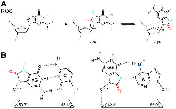
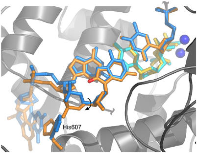
Accurate DNA replication involves polymerases with high nucleotide selectivity and proofreading activity. We show here why both fidelity mechanisms fail when normally accurate T7 DNA polymerase bypasses the common oxidative lesion 8-oxo-7, 8-dihydro-2'-deoxyguanosine (8oG). The crystal structure of the polymerase with 8oG templating dC insertion shows that the O8 oxygen is tolerated by strong kinking of the DNA template. A model of a corresponding structure with dATP predicts steric and electrostatic clashes that would reduce but not eliminate insertion of dA. The structure of a postinsertional complex shows 8oG(syn).dA (anti) in a Hoogsteen-like base pair at the 3' terminus, and polymerase interactions with the minor groove surface of the mismatch that mimic those with undamaged, matched base pairs. This explains why translesion synthesis is permitted without proofreading of an 8oG.dA mismatch, thus providing insight into the high mutagenic potential of 8oG.


Figures

Figure 1
Figure 1
The dual coding potential of 8oG is explained by its unique chemical characteristics and physical properties. (A) Oxidation of guanosine by reactive oxygen species (ROS) leads to the formation of 8oG, which is distinguished from guanosine by the O8 group (colored red) and the protonation of nitrogen N7 (colored blue). Nucleosides have free rotation at their glycosidic bond (arrow); thus, they exist in equilibrium between syn and anti conformations. Whereas guanosine predominates in an anti conformation, the syn conformation is favored in 8oG, presumably due to the unfavorable steric interaction between the O8 group and the ribose O4 oxygen. (B) 8oG forms both Watson–Crick and Hoogsteen base pairs (Lipscomb et al, 1995; McAuley-Hecht et al, 1994). An 8oG (anti)·dC base pair (left) is structurally identical to a canonical dG·dC base pair, in that the anti conformation of the 8oG allows for hydrogen bonding between the Watson–Crick faces of both bases, and that the C1′–C1′ distance and the minor groove hydrogen bond donors and acceptors are conserved. The protonated N7 and O8 groups project into the major groove. The 8oG (syn)·dA base pair (right) is formed from the Hoogsteen face of 8oG in syn with the Watson–Crick face of adenine in anti. In this mismatch, the N1, N2 and O6 atoms of the 8oG (syn) project into the major grove and the O8 groups project into the minor groove of the base pair.
Figure 2
Figure 2
T7 DNA polymerase bypass of an 8oG lesion. Primer extension reactions were performed with exo (left) and wild-type (right) T7 DNA polymerase with undamaged guanine (G) and 8-oxoguanine (8oG) in comparison to controls containing no enzyme. The images shown are for 3 min incubations of reaction mixtures containing 200- to 400-fold excess of DNA over polymerase. The most intense band in each lane is unreacted primer, at least 80% of which remains unextended for all efficiency reactions performed in this study. The location of 8oG within the template strand is as indicated and enhanced images of products using 8oG are shown to the right of the boxed images. The probability of insertion at each template site, listed in percent to the right of each lane, is an average of 7–16 determinations and is calculated as described previously (Kokoska et al, 2003).
Figure 3
Figure 3
Structural rationale for deoxycytidine insertion opposite 8oG. The σA-weighted FoFc annealed omit electron density maps, contoured at 3σ, are shown superimposed onto the refined models of incoming ddCTP across from template guanosine (A) and template 8oG (B). In the 8oG·dCTP insertion complex, Lys536 (gold bonds) moves 3 Å relative to its position in the native dG·dCTP complex and forms a hydrogen bond with the O8 group. The positions of the Mg2+ ions (yellow spheres) are similar to previously determined T7 DNA polymerase closed complexes. (C) Superposition of ternary complexes of T7 DNA polymerase and Pol β with G and 8oG at the templating position. The conformations of an 8oG·dCTP insertion complex (orange) and native G·dCTP insertion complex (yellow) are almost identical. A superposition between both complexes reveals the conservation of a DNA kink of the 5′ phosphate backbone at the templating base. A superposition of a dG·dCTP insertion of pol β (green) shows that the kink is not as pronounced and a steric clash between the 5′ phosphate and the O8 group is prone to occur. However, the structure of pol β in an 8oG·dCTP insertion complex (blue) reveals that pol β is able to incorporate dCTP across from the lesion because of a local conformational change of the DNA backbone.
Figure 4
Figure 4
Comparison of an open 8oG complex and a closed T·ddATP insertion complex. The open 8oG complex (red) and a dT·dATP insertion complex (gray) were superimposed using Cα atoms. The proteins are depicted as cylinders and the DNA as sticks. Both structures are largely similar but they specifically differ in the orientation adopted by their fingers subdomains. In the closed structure, α-helices O and O1 pack against the incoming ddATP (blue) and the template thymine, respectively. In the open structure, the fingers move outwards from the palm subdomain, as shown by the ∼45° rotation of the O and O1 helices relative to the closed conformation. Residue Tyr530, which moves to the position that would correspond to the templating base of the closed complex, has been omitted for clarity. The templating 8oG, the 5′ template strand, and residues 532–536 located at the junction between α helices O and O1 are disordered in the open complex. No interpretable electron density is observed for the metal ions or incoming nucleotide.
Figure 5
Figure 5
Model of a catalytic complex of dATP paired with 8oG. Modeling an 8oG(syn)·dATP pair from the 8oG·dA postinsertion complex into the polymerase active site shows that the amino acids from the O and O1 helices hinder the incorporation of dATP (yellow carbons) across from an 8oG lesion (cyan carbons). van der Waals surfaces are shown for protein side chains (orange) and 8oG (white). Unfavorable interactions between the 8oG and protein atoms are represented as red patches on the van der Waals surfaces. The N1 and N2 atoms of 8oG make detrimental electrostatic interactions with the ɛ and δ amino groups of Lys536 and the γ carbon of Ile540. The O8 group also makes steric clashes with residue Tyr530.
Figure 6
Figure 6
Structural rationale for elongation from an 8oG lesion. The 8oG (anti)·dC (anti) postinsertion complexes are shown in panels A, C and E. 8oG (syn)·dA (anti) postinsertion complexes are shown in panels B, D and F. (A, B) The σA-weighted FoFc annealed omit electron density maps, contoured at 3σ, for the elongated 8oG·dCMP (A) and 8oG·dAMP (B) structures are shown against the refined models. The topology of the major groove is highlighted with dashed arcs. In the 8oG (syn)·dA base pair, the adenine N1, N2 and O6 atoms protrude into the major groove and the O8 group is located at the minor groove. (C, D) Molecular surface representations of the DNA in the elongation complexes. The major groove surfaces corresponding to the 8oG atoms are colored red and the Mg2+ ions are represented as green spheres. The major groove surface of the 8oG·dC postinsertion complex (C) is similar to that of a canonical dG·dC base pair, whereas 8oG (syn) paired with dA forms a bump in the major groove (D). However, no steric clashes are observed between the polymerase and the bulky 8oG·dA pair. (E, F) Superpositions between 8oG postinsertion complexes and normal DNA. (E) A superposition between the 8oG·dC postinsertion complex (blue) and a normal postinsertion dA·dT complex (yellow) shows that the minor groove groups are positioned in an identical manner between the lesioned and the unmodified purine·pyrimidine base pairs. The acceptors are recognized by residues Gln615 and Arg429, which hydrogen bond with N7 of the 8oG base and O2 of the incorporated dC. The phosphate backbone of the 8oG rotates by 90° about its γ torsion angle in comparison to an unlesioned base, in order to avoid a steric clash with the O8 group. (F) A superposition between an 8oG·dA postinsertion complex (green) and a postinsertion dT·dA complex (orange) shows that the minor groove is identical between the lesioned and the unmodified pyrimidine·purine base pairs. Residues Gln615 and Arg429 form hydrogen bonds with the O8 group of 8oG and with the N3 group of the incorporated dA. The O8 group mimics the O2 group of a thymidine.
Figure 7
Figure 7
Bias against elongation from 8oG·dC. A superposition between the 8oG (anti)·dC (anti) postinsertion complex (blue) and a dG·dCTP insertion complex (orange) shows that a rotation around the C5′–C4′ bond of the 8oG phosphate backbone is necessary to avoid a steric clash with oxygen O8. This movement is translated to the adjacent nucleotide and it shifts the position of the sugar moiety of the templating strand by ∼1.5 Å in comparison to an unmodified template. As the C5′–C4′ bond of an 8oG (syn)·dA (anti) postinsertion complex is similar to a native dG·dCTP insertion complex, the subtle shift in the sugar moiety of an 8oG (anti)·dC (anti) pair may decrease the catalytic efficiency of nucleotide insertion, explaining the preference for elongation of a mismatch versus a correct pair. The imidazole ring of His607 tracks along with the 5′ phosphate for both elongation complexes as is observed for the original ternary complex, supporting its role in positioning the template.

References

    1. Beckman KB, Ames BN (1997) Oxidative decay of DNA. J Biol Chem 272: 19633–19636 - PubMed
    1. Boudsocq F, Kokoska RJ, Plosky BS, Vaisman A, Ling H, Kunkel TA, Yang W, Woodgate R (2004) Investigating the role of the little finger domain of Y-family DNA polymerases in low-fidelity synthesis and translesion replication. J Biol Chem, (in press) - PubMed
    1. Braithwaite DK, Ito J (1993) Compilation, alignment, and phylogenetic relationships of DNA polymerases. Nucleic Acids Res 21: 787–802 - PMC - PubMed
    1. Brautigam CA, Steitz TA (1998) Structural and functional insights provided by crystal structures of DNA polymerases and their substrate complexes. Curr Opin Struct Biol 8: 54–63 - PubMed
    1. Brunger AT, Adams PD, Clore GM, DeLano WL, Gros P, Grosse-Kunstleve RW, Jiang JS, Kuszewski J, Nilges M, Pannu NS, Read RJ, Rice LM, Simonson T, Warren GL (1998) Crystallography & NMR system: a new software suite for macromolecular structure determination. Acta Crystallogr D 54: 905–921 - PubMed

Publication types

MeSH terms

LinkOut - more resources