

Genome-wide array comparative genomic hybridization analysis reveals distinct amplifications in osteosarcoma
- PMID: 15298715
- PMCID: PMC514550
- DOI: 10.1186/1471-2407-4-45
Genome-wide array comparative genomic hybridization analysis reveals distinct amplifications in osteosarcoma
Abstract
Background: Osteosarcoma is a highly malignant bone neoplasm of children and young adults. It is characterized by extremely complex karyotypes and high frequency of chromosomal amplifications. Currently, only the histological response (degree of necrosis) to therapy represent gold standard for predicting the outcome in a patient with non-metastatic osteosarcoma at the time of definitive surgery. Patients with lower degree of necrosis have a higher risk of relapse and poor outcome even after chemotherapy and complete resection of the primary tumor. Therefore, a better understanding of the underlying molecular genetic events leading to tumor initiation and progression could result in the identification of potential diagnostic and therapeutic targets.
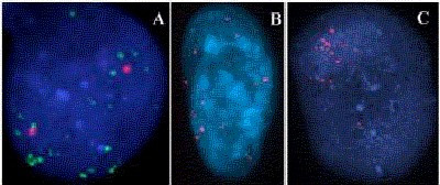
Methods: We used a genome-wide screening method - array based comparative genomic hybridization (array-CGH) to identify DNA copy number changes in 48 patients with osteosarcoma. We applied fluorescence in situ hybridization (FISH) to validate some of amplified clones in this study.
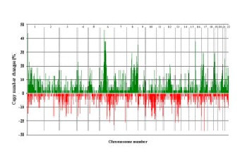
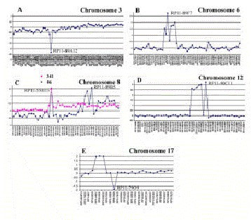
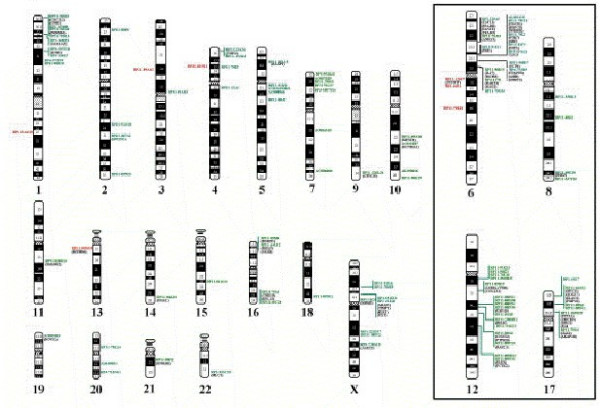
Results: Clones showing gains (79%) were more frequent than losses (66%). High-level amplifications and homozygous deletions constitute 28.6% and 3.8% of tumor genome respectively. High-level amplifications were present in 238 clones, of which about 37% of them showed recurrent amplification. Most frequently amplified clones were mapped to 1p36.32 (PRDM16), 6p21.1 (CDC5L, HSPCB, NFKBIE), 8q24, 12q14.3 (IFNG), 16p13 (MGRN1), and 17p11.2 (PMP22 MYCD, SOX1,ELAC27). We validated some of the amplified clones by FISH from 6p12-p21, 8q23-q24, and 17p11.2 amplicons. Homozygous deletions were noted for 32 clones and only 7 clones showed in more than one case. These 7 clones were mapped to 1q25.1 (4 cases), 3p14.1 (4 cases), 13q12.2 (2 cases), 4p15.1 (2 cases), 6q12 (2 cases), 6q12 (2 cases) and 6q16.3 (2 cases).
Conclusions: This study clearly demonstrates the utility of array CGH in defining high-resolution DNA copy number changes and refining amplifications. The resolution of array CGH technology combined with human genome database suggested the possible target genes present in the gained or lost clones.
Figures
References
-
- Huvos A. In: Bone Tumors: Diagnosis, Treatment and Prognosis, Huvos A, editor. Philadelphia, WB Saunders; 1991.
-
- Provisor AJ, Ettinger LJ, Nachman JB, Krailo MD, Makley JT, Yunis EJ, Huvos AG, Betcher DL, Baum ES, Kisker CT, Miser JS. Treatment of nonmetastatic osteosarcoma of the extremity with preoperative and postoperative chemotherapy: a report from the children's cancer group. J Clin Oncol. 1997;15:76–84. - PubMed
-
- Tarkkanen M, Karhu R, Kallioniemi A, Elomaa I, Kivioja AH, Nevalainen J, Bohling T, Karaharju E, Hyytinen E, Knuutila S. Gains and losses of DNA sequences in osteosarcomas by comparative genome hybridization. Cancer Res. 1995;55:1334–1338. - PubMed
-
- Tarkkanen M, Elomaa I, Blomqvist C, Kivioja AH, Kellokumpu-Lehtinen P, Bohling T, Valle J, Knuutila S. DNA sequence copy number increase at 8q: a potential new prognostic marker in high-grade osteosarcoma. Int J Cancer. 1999;84:114–121. doi: 10.1002/(SICI)1097-0215(19990420)84:2<114::AID-IJC4>3.0.CO;2-Q. - DOI - PubMed
Publication types
MeSH terms
Grants and funding
LinkOut - more resources
Full Text Sources
Other Literature Sources
Medical
Miscellaneous
