. 2004 Aug 6;32(14):4224-36.
doi: 10.1093/nar/gkh752. Print 2004.

hnRNP H binding at the 5' splice site correlates with the pathological effect of two intronic mutations in the NF-1 and TSHbeta genes

Affiliations

hnRNP H binding at the 5' splice site correlates with the pathological effect of two intronic mutations in the NF-1 and TSHbeta genes

Emanuele Buratti et al. Nucleic Acids Res. .

Abstract

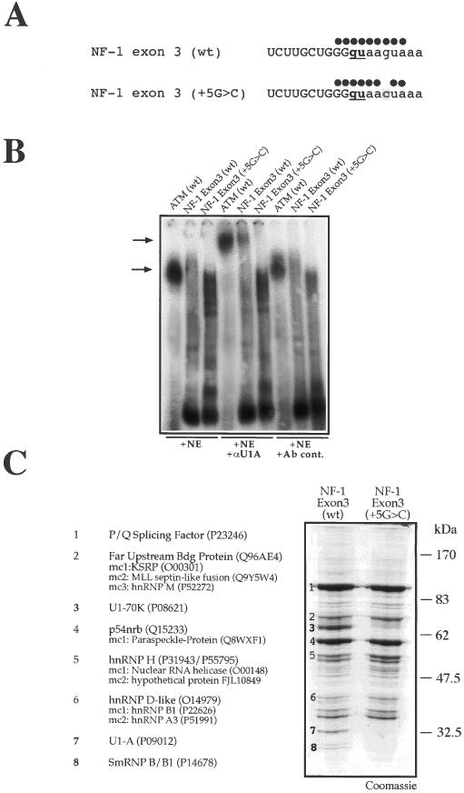
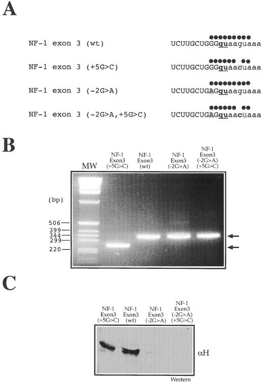
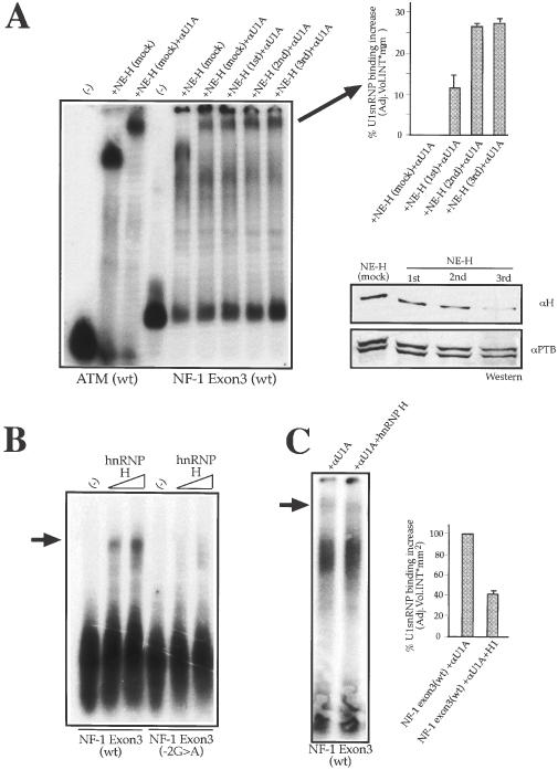
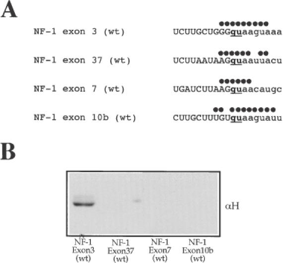
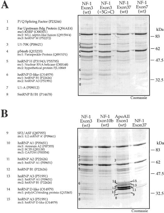
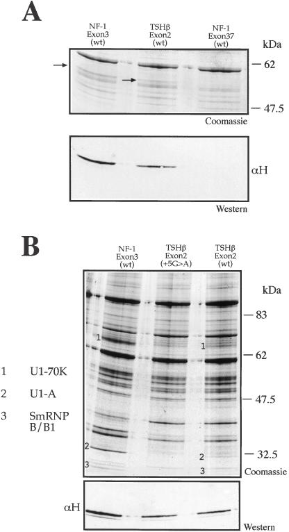
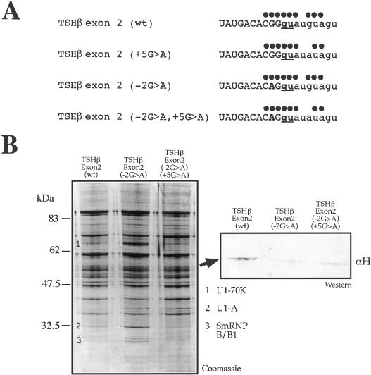
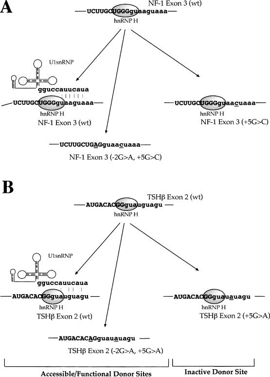
We have recently reported a disease-causing substitution (+5G > C) at the donor site of NF-1 exon 3 that produces its skipping. We have now studied in detail the splicing mechanism involved in analyzing RNA-protein complexes at several 5' splice sites. Characteristic protein patterns were observed by pulldown and band-shift/super-shift analysis. Here, we show that hnRNP H binds specifically to the wild-type GGGgu donor sequence of the NF-1 exon 3. Depletion analyses shows that this protein restricts the accessibility of U1 small nuclear ribonucleoprotein (U1snRNA) to the donor site. In this context, the +5G > C mutation abolishes both U1snRNP base pairing and the 5' splice site (5'ss) function. However, exon recognition in the mutant can be rescued by disrupting the binding of hnRNP H, demonstrating that this protein enhances the effects of the +5G > C substitution. Significantly, a similar situation was found for a second disease-causing +5G > A substitution in the 5'ss of TSHbeta exon 2, which harbors a GGgu donor sequence. Thus, the reason why similar nucleotide substitutions can be either neutral or very disruptive of splicing function can be explained by the presence of specific binding signatures depending on local contexts.


Figures

Figure 1
Figure 1
U1snRNP binds to wild type but not mutated NF-1 exon 3. (A) Schematic representation of the U1snRNA complementarity with NF-1 exon 3 wild-type (wt) and containing the NF-1 exon 3 +5G > C substitution. Full circles above each 5′ splice site sequence represent base pair matches with U1snRNA. (B) Super-shift analysis of nuclear complexes using a monoclonal antibody against U1-A protein (αU1A). The different RNAs are incubated with nuclear extract (NE) either alone or in the presence of an anti-U1A mAb (αU1A) or a control antibody (Ab cont.). The shifted complexes are indicated by the lower arrow while super-shifted αU1A-U1snRNP–RNA complexes by the upper arrow. As positive control a 20mer synthetic ribonucleotide (ATM WT) that has been previously shown to be a high affinity U1snRNP binder is used. (C) Coomassie Blue staining of a pulldown assay using adipic acid dehydrazide beads coated with NF-1 exon 3 (wt) and NF-1 exon 3 (+5G > C) RNAs following incubation with HeLa nuclear extract. In the lane from the exon 3 (wt) the numbers in boldface indicate the 70, 32.5 and 25 kDa protein bands that are absent in the NF-1 exon 3 (+5G > C) lane and which belong to the U1snRNP complex. Major protein identities are reported on the left including their Swiss-Prot Accession number. When present, minor protein components (mc) are also reported in the order of occurrence.
Figure 2
Figure 2
Removal of putative hnRNP H binding site results in recovery of splicing activity. (A) Nucleotide sequences around the 5′ss of NF-1 exon 3 (wt) and NF-1 exon 3 (+5G > C) following the introduction of the −2G > A substitution. Full circles above each 5′ splice site sequence represent base pair matches with U1snRNA. (B) Effect of the −2G > A substitution both in NF-1 exon 3 (wt) and in the NF-1 exon 3 (+5G > C) mutant following introduction in the EDB minigene and transfection in HeLa cells. Two products (see arrows) are seen after RNA extraction and RT–PCR analysis on agarose gel electrophoresis: the upper band (323 nt) includes exon 3 while the lower band (239 nt) lacks this exon. Note that the −2G > A mutation completely rescues NF-1 exon 3 splicing inhibition in the mutant bearing the +5G > C substitution. (C) Pulldown analysis of NF-1 exon 3 (−2G > A) and NF-1 exon 3 (−2G > A, +5G > C) RNAs followed by western blot analysis to determine the extent of hnRNP H binding to each RNA.
Figure 3
Figure 3
Depletion/addition of hnRNP H from the nuclear extract can affect U1snRNP binding to NF-1 exon 3 (wt) donor site. (A) Left panel shows a super-shift analysis of NF-1 exon 3 (wt) donor site RNA using a monoclonal antibody against U1snRNP U1-A protein (αU1A). The exon 3 (wt) RNA was incubated with a mock-depleted nuclear extract NE (mock) and with equal amounts of nuclear extract treated with a polyclonal antibody against hnRNP H (αH) in three successive rounds of depletion: NE-H (first), NE-H (second), and NE-H (third). As control, the ATM (wt) RNA is incubated with NE (mock) in the absence or presence of (αU1A). The super-shifted αU1A-U1snRNP–RNA complexes were quantified using the Quantity One program (Bio-Rad) (upper right panel). SD bars are shown. (A) Lower right panel shows the levels of hnRNP H from the nuclear extract with the three successive round of depletion with αH. As control, a polyclonal antibody against Polypyrimidine Tract Binding protein (αPTB) was used to test each depleted extract for aspecific depletion. (B) Shows a band-shift experiment using labeled NF-1 exon 3 (wt) and NF-1 exon 3 (−2G > A) RNAs incubated with increasing quantities of recombinant hnRNP H (500 ng and 1 μg respectively). The arrow on the left indicates the hnRNP H–RNA complex. (C) Shows the results of adding 1 μg of recombinant hnRNP H to NF-1 exon 3 (wt) RNA in the presence of nuclear extract and an antibody against U1snRNP (αU1). The arrow indicates the super-shifted αU1A–U1snRNP–RNA complex which was quantified using the Quantity One program (Bio-Rad). SD bars are shown.
Figure 4
Figure 4
U1snRNP and hnRNP H do not bind to NF-1 exon 37, 7 and 10b donor sites carrying substitutions in the +5 or −1 position. (A) Nucleotide sequences around the 5′ss of NF-1 exon 3 (wt), NF-1 exon 7 (wt), NF-1 exon 37 (wt) and NF-1 exon 10b (wt) (see Table 1 for complete sequence) showing the region of complementarity with U1snRNA (indicated by black dots). (B) Western blot of pulldown analyses of all these RNAs to determine the eventual presence of hnRNP H.
Figure 5
Figure 5
Ribonucleoprotein complexes in the 5′ss of NF-1 and Apo AII donor sites. (A) Coomassie Blue staining of a pulldown assay using beads coated with NF-1 exon 3 (wt), NF-1 exon 3 (+5G > C), NF-1 exon 7 (wt), and NF-1 exon 37 (wt) RNAs following incubation with HeLa nuclear extract. The numbers on the NF-1 exon 3 (wt) lane indicate the sequenced protein bands. The identity of each numbered band is shown to the left of the gel. (B) Coomassie Blue staining of a pulldown assay using beads derivatized with NF-1 exon 3 (wt), NF-1 exon 10b (wt) and Apo AII exon 1 (wt), and a control pulldown using exonic sequence from NF-1 exon 37 lacking a 5′ splice site. The numbers on the NF-1 exon 3 (wt) lane indicate the sequenced protein bands. On the left of the gel the identity of each band together with its Swiss-Prot Accession number is reported. Minor components (mc), when present, are also reported (in decreasing order of detection).
Figure 6
Figure 6
hnRNP H binds to the donor site of TSHβ exon 2 type. (A) Coomassie and western blot analysis of a pulldown assay using NF-1 exon 3 (wt), TSHβ exon 2 (wt) and NF-1 exon 37 (wt) RNAs. The arrows indicate the protein signatures corresponding to hnRNP H in the Coomassie gel. These signatures were confirmed by western blot analysis using an anti-hnRNP H antibody (αH) in the lower panel. (B) Coomassie Blue staining of a pulldown assay using adipic acid dehydrazide beads coated with NF-1 exon 3 (wt), TSHβ exon 2 (+5G > A), and TSHβ exon 2 (wt) RNAs following incubation with HeLa nuclear extract. The numbers indicate the U1snRNP components previously identified: U1-70K (1), U1-A (2) and SmRNP B/B1 (3) and the lower panel contains a western blot performed on the Coomassie gel to determine hnRNP H binding to the different RNAs.
Figure 7
Figure 7
Removal of putative hnRNP H binding site in TSHβ exon 2 (wt) and TSHβ exon 2 (+5G > A). (A) Nucleotide sequences around the 5′ss of TSHβ exon 2 (wt) and TSHβ exon 2 (+5G > A) following the introduction of the −2G > A substitution. Dots above each 5′ splice site sequence represent base pair matches with U1snRNA. (B) Pulldown and western blot analysis performed on TSHβ exon 2 (wt), TSHβ exon 2 (−2G > A), and TSHβ exon 2 (−2G > A, +5G > A) RNAs to determine the extent of U1snRNP binding to each RNA. The numbers indicate the U1snRNP components previously identified: U1-70K (1), U1-A (2) and SmRNP B/B1 (3). The Coomassie gel was used in western blot analysis (panel on the right) to determine the effects of the −2G > A substitution on binding of hnRNP H to these RNAs.
Figure 8
Figure 8
Model of donor site inhibition by hnRNP H. The diagrams represent the proposed model of splicing inhibition mediated by hnRNP H binding in correspondence to the NF-1 exon 3 (wt) donor site when the +5G > C mutation occurs and in the TSHβ exon 2 donor site following the +5G > A substitution. Both wild-type donor site sequences are capable of binding U1snRNP stably owing to the complementarity still present toward the U1snRNA sequence even in the presence of hnRNP H binding near the central GU dinucleotide. Donor site recognition can be abolished by the introduction of the +5G > C or +5G > A mutations which in both cases do not affect hnRNP H binding but lower U1snRNA complementarity with the donor site below a critical threshold. Finally, removal of hnRNP H binding site is not sufficient to recover U1snRNP binding (owing to the presence of the +5 substitutions) but is sufficient to allow recognition of the donor site by the splicing machinery.

References

    1. Burge C.B., Tuschl,T. and Sharp,P.A. (1999) Splicing of Precursors to mRNAs by the Spliceosome. Cold Spring Laboratory Harbor Press, Cold Spring Harbor, NY.
    1. Mount S.M., Pettersson,I., Hinterberger,M., Karmas,A. and Steitz,J.A. (1983) The U1 small nuclear RNA–protein complex selectively binds a 5′ splice site in vitro. Cell, 33, 509–518. - PubMed
    1. Stark H., Dube,P., Luhrmann,R. and Kastner,B. (2001) Arrangement of RNA and proteins in the spliceosomal U1 small nuclear ribonucleoprotein particle. Nature, 409, 539–542. - PubMed
    1. Zhang M.Q. (1998) Statistical features of human exons and their flanking regions. Hum. Mol. Genet., 7, 919–932. - PubMed
    1. Michaud S. and Reed,R. (1991) An ATP-independent complex commits pre-mRNA to the mammalian spliceosome assembly pathway. Genes Dev., 5, 2534–2546. - PubMed

Publication types

MeSH terms

Substances