Comparative Study
. 2004 Aug;135(4):2055-67.
doi: 10.1104/pp.104.040873. Epub 2004 Aug 6.

Differential antifungal and calcium channel-blocking activity among structurally related plant defensins

Affiliations
Comparative Study

Differential antifungal and calcium channel-blocking activity among structurally related plant defensins

Robert G Spelbrink et al. Plant Physiol. 2004 Aug.

Abstract

Plant defensins are a family of small Cys-rich antifungal proteins that play important roles in plant defense against invading fungi. Structures of several plant defensins share a Cys-stabilized alpha/beta-motif. Structural determinants in plant defensins that govern their antifungal activity and the mechanisms by which they inhibit fungal growth remain unclear. Alfalfa (Medicago sativa) seed defensin, MsDef1, strongly inhibits the growth of Fusarium graminearum in vitro, and its antifungal activity is markedly reduced in the presence of Ca(2+). By contrast, MtDef2 from Medicago truncatula, which shares 65% amino acid sequence identity with MsDef1, lacks antifungal activity against F. graminearum. Characterization of the in vitro antifungal activity of the chimeras containing portions of the MsDef1 and MtDef2 proteins shows that the major determinants of antifungal activity reside in the carboxy-terminal region (amino acids 31-45) of MsDef1. We further define the active site by demonstrating that the Arg at position 38 of MsDef1 is critical for its antifungal activity. Furthermore, we have found for the first time, to our knowledge, that MsDef1 blocks the mammalian L-type Ca(2+) channel in a manner akin to a virally encoded and structurally unrelated antifungal toxin KP4 from Ustilago maydis, whereas structurally similar MtDef2 and the radish (Raphanus sativus) seed defensin Rs-AFP2 fail to block the L-type Ca(2+) channel. From these results, we speculate that the two unrelated antifungal proteins, KP4 and MsDef1, have evolutionarily converged upon the same molecular target, whereas the two structurally related antifungal plant defensins, MtDef2 and Rs-AFP2, have diverged to attack different targets in fungi.


Figures

Figure 1.
Figure 1.
Sequence comparison of Rs-AFP2 and Medicago defensins. Significant differences in residues between the two Medicago defensins are shown in boldface. The charge of each protein is shown in parentheses. Symbols represent approximate positions of the predicted α-helix (spiral) and β-sheets (arrows).
Figure 2.
Figure 2.
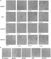
Comparison between the effects of KP4, MsDef1, Rs-AFP2, and MtDef2 on F. graminearum. A, MsDef1, KP4, and Rs-AFP2 all exhibit a dose-dependent inhibition of hyphae. Assays were conducted in low ionic strength synthetic fungal media inoculated with fungal spores. Micrographs prepared after a 16-h incubation. B, Hyperbranching effect on Fusarium. In vitro assays were prepared in the same manner as described in A, with 25 μg mL−1 of antifungal protein. The average number of hyphal buds per germline was determined by counting 50 germlings after 9 h of incubation. There is a marked difference in the number of buds among the proteins tested, with MsDef1 and KP4 inducing the most buds, Rs-AFP2 causing a less extreme effect, and MtDef2 causing no hyperbranching.
Figure 3.
Figure 3.
Antifungal activity of the chimeric Def1/Def2 defensins on F. graminearum. MsDef1 and MtDef2 were divided into three regions of similar length, and chimeric proteins corresponding to all six possible combinations, termed Def1-2C1 through Def1-2C6, were obtained by expressing the synthetic genes encoding these proteins using a P. pastoris expression system. All six chimeric proteins were screened for antifungal activity against F. graminearum and N. crassa (data not shown), along with MsDef1 and MtDef2 proteins. Antifungal activity was assessed by measuring both hyphal growth inhibition after 16 h of exposure to the proteins and determining the degree of hyperbranching after 9 h of exposure to the proteins (Table II).
Figure 4.
Figure 4.
Arg-38 of MsDef1 is important for antifungal activity. Spores of both fungi were allowed to germinate and grow in synthetic fungal media supplemented with 12 μg mL−1 of the indicated defensin for N. crassa and 25 μg mL−1 defensins for F. graminearum. Growth inhibition and hyperbranching were determined as described in “Materials and Methods.”
Figure 5.
Figure 5.
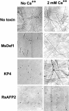
Abrogation of the antifungal activity of MsDef1 and KP4 by Ca2+. Shown here is an example of one of the tested metals and concentrations; 50 μg mL−1 KP4 and 5 μg mL−1 MsDef1 and Rs-AFP2 were used in this experiment.
Figure 6.
Figure 6.
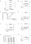
MsDef1, but not Rs-AFP2 or MtDef2, selectively blocks L-type Ca2+ channels. A, Time dependency of the MsDef1 block of Cav1.2 channels. As shown here, 10 μm MsDef1 blocks approximately 90% of the Ca2+ current, with the maximum inhibition occurring after exposing the cells to the defensin for approximately 13 min. B, Block by 2 μm MsDef1 developed slowly, over several minutes, such that 0.56% ± 0.03% of current remained at equilibrium (n = 3). C, Current-voltage relationship of Cav1.2 in the presence and absence of 2 μm MsDef1. Cells expressing Cav1.2, as described above, were held at −60 mV and depolarized to the indicated voltage for 100 ms before (black circles) or after (white circles) equilibration in 2 μm MsDef1. While MsDef1 decreases peak current, it does not appreciably shift the current-voltage relationship of Cav1.2. D and E, MsDef1 was applied to the non-L-type channels Cav2.1 and Cav 2.3, respectively, as in B, except that the holding potential was −80 mV for Cav 2.1 and −100 mV for Cav 2.3. No inhibition of either Cav2.1 or Cav 2.3 by MsDef1 was detected. F, Whole-cell voltage clamp of tsA-201 cells expressing Cav1.2 channels, as in B, before (control) and several minutes after the application of 10 μm Rs-AFP2. G, The fraction of control current remaining after several minutes of perfusion with Rs-AFP2 in tsA-201 cells expressing Cav1.2, Cav2.1, or Cav2.3 channels. Ba2+ currents in these channels were elicited using the protocol described in B and D. The defensin Rs-AFP2 did not inhibit current conducted by any of these voltage-gated Ca2+ channels (values are means ± se; n = 3). H, L-type Cav1.2 channels are not blocked by MtDef2. The cell was held at −60 mm and pulsed to +10 mV before (control) or 5 min after initiation of perfusion with 10 μm MtDef2.
Figure 7.
Figure 7.
Three-dimensional structures of the plant defensin Rs-AFP1 (1AYJ; Fant et al., 1998), the scorpion toxin AaHII (1PTX; Housset et al., 1994), and the fungal toxin KP4 (1KPT; Gu et al., 1995). In all three figures, the Cys side chains are represented in ball and stick, while the Arg and Lys residues are shown as blue stick models. In the diagram of Rs-AFP1, the colors of the secondary structural elements correspond to the regions selected for MsDef1 hybrid analysis. Orange, purple, and green indicate the N-terminal (residues 1–15), middle (residues 16–30), and C-terminal (residues 31–45) portions, respectively. For scorpion toxin and KP4, the ribbon diagrams are colored in a gradient from red to purple as the protein is traced from the N to the C termini.

References

    1. Almeida MS, Cabral KM, Kurtenbach E, Almeida FC, Valente AP (2002) Solution structure of Pisum sativum defensin 1 by high resolution NMR: plant defensins, identical backbone with different mechanisms of action. J Mol Biol 315: 749–757 - PubMed
    1. Bloch C, Richardson M (1991) A new family of small (5 kDa) protein inhibitors of insect alpha-amylases from seeds of Sorghum (Sorghum bicolor (L) Moench) have sequence homologies with wheat gamma-purothionins. FEBS Lett 279: 101–105 - PubMed
    1. Bontems F, Roumestand C, Boyot P, Gilquin B, Doljansky Y, Menez A, Toma F (1991) Three-dimensional structure of natural charybdotoxin in aqueous solution by 1H-NMR. Charybdotoxin possesses a structural motif found in other scorpion toxins. Eur J Biochem 196: 19–28 - PubMed
    1. Borkovich KA, Alex LA, Yarden O, Freitag M, Turner GE, Read ND, Seiler S, Bell-Pedersen D, Paietta J, Plesofsky N, et al (2004) Lessons from the genome sequence of Neurospora crassa: tracing the path from genomic blueprint to multicellular organism. Microbiol Mol Biol Rev 68: 1–108 (table of contents) - PMC - PubMed
    1. Broekaert WF, Terras FR, Cammue BP, Osborn RW (1995) Plant defensins: novel antimicrobial peptides as components of the host defense system. Plant Physiol 108: 1353–1358 - PMC - PubMed

Publication types

Associated data

LinkOut - more resources