

Detection of BCL2-IGH rearrangement on paraffin-embedded tissue sections obtained from a small submucosal tumor of the rectum in a patient with recurrent follicular lymphoma
- PMID: 15300917
- PMCID: PMC4572174
- DOI: 10.3748/wjg.v10.i17.2602
Detection of BCL2-IGH rearrangement on paraffin-embedded tissue sections obtained from a small submucosal tumor of the rectum in a patient with recurrent follicular lymphoma
Abstract
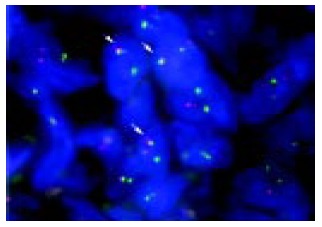
A 59-year-old woman was admitted to our hospital because of recurrent follicular lymphoma (FL). Colonoscopic examination revealed a rectal submucosal tumor (SMT) without any erosions and ulcers. In this patient, it was difficult to distinguish non-Hodgkin's lymphoma (NHL) invasion from other disorders of the colon including carcinoid tumor merely based on endoscopic findings. Histopathologic and immunohistochemical studies on biopsy specimens showed an infiltration of atypical lymphocytes that were positive for CD20 and BCL2 but negative for UCHL-1. Fluorescence in situ hybridization on paraffin-embedded tissue sections (T-FISH) identified a translocation of BCL2 with IGH gene. Based on these findings, the tumor was defined as an invasion of FL. T-FISH method is useful for the detection of a monoclonality of atypical lymphocytes in an SMT of the gastrointestinal tract, and particularly for the detection of chromosomal translocations specific to lymphoma subtypes.
Copyright 2004 The WJG Press ISSN
Figures


Similar articles
-
Fluorescence in situ hybridization detection of chromosome IGH/BCL2 translocations from paraffin-embedded tissue: evaluation in follicular lymphoma.Int J Hematol. 2003 Aug;78(2):154-9. doi: 10.1007/BF02983385. Int J Hematol. 2003. PMID: 12953811
-
Detection of t(14;18) in follicular lymphoma by dual-color fluorescence in situ hybridization on paraffin-embedded tissue sections.Cancer Genet Cytogenet. 2004 Apr 1;150(1):22-6. doi: 10.1016/j.cancergencyto.2003.08.008. Cancer Genet Cytogenet. 2004. PMID: 15041219
-
FISH is superior to PCR in detecting t(14;18)(q32;q21)-IgH/bcl-2 in follicular lymphoma using paraffin-embedded tissue samples.Am J Clin Pathol. 2005 Sep;124(3):421-9. doi: 10.1309/BLH8-MMK8-5UBQ-4K6R. Am J Clin Pathol. 2005. PMID: 16191511
-
Utility of immunohistochemistry with an antibody against MYC at the initial diagnosis of follicular lymphoma, grade 3A, for predicting a more aggressive clinical course: a case report and review of the literature.Int J Clin Exp Pathol. 2015 Jun 1;8(6):7559-64. eCollection 2015. Int J Clin Exp Pathol. 2015. PMID: 26261669 Free PMC article. Review.
-
Immunophenotypic and cytogenetic findings of B-lymphoblastic leukemia/lymphoma associated with combined IGH/BCL2 and MYC rearrangement.Cytometry B Clin Cytom. 2017 Jul;92(4):310-314. doi: 10.1002/cyto.b.21334. Epub 2016 Feb 5. Cytometry B Clin Cytom. 2017. PMID: 26517296 Review.
Cited by
-
Gastrointestinal follicular lymphoma: review of the literature.J Gastroenterol. 2010 Apr;45(4):370-88. doi: 10.1007/s00535-009-0182-z. Epub 2010 Jan 20. J Gastroenterol. 2010. PMID: 20084529 Review.
-
Trisomy 3 may predict a poor response of gastric MALT lymphoma to Helicobacter pylori eradication therapy.World J Gastroenterol. 2005 Jan 7;11(1):89-93. doi: 10.3748/wjg.v11.i1.89. World J Gastroenterol. 2005. PMID: 15609403 Free PMC article.
-
Diagnosis of follicular lymphoma of the gastrointestinal tract: A better initial diagnostic workup.World J Gastroenterol. 2016 Jan 28;22(4):1674-83. doi: 10.3748/wjg.v22.i4.1674. World J Gastroenterol. 2016. PMID: 26819532 Free PMC article. Review.
References
-
- Frazee RC, Roberts J. Gastric lymphoma treatment. Medical versus surgical. Surg Clin North Am. 1992;72:423–431. - PubMed
-
- Radaszkiewicz T, Dragosics B, Bauer P. Gastrointestinal malignant lymphomas of the mucosa-associated lymphoid tissue: factors relevant to prognosis. Gastroenterology. 1992;102:1628–1638. - PubMed
-
- Hansen PB, Vogt KC, Skov RL, Pedersen-Bjergaard U, Jacobsen M, Ralfkiaer E. Primary gastrointestinal non-Hodgkin's lymphoma in adults: a population-based clinical and histopathologic study. J Intern Med. 1998;244:71–78. - PubMed
-
- Crump M, Gospodarowicz M, Shepherd FA. Lymphoma of the gastrointestinal tract. Semin Oncol. 1999;26:324–337. - PubMed
Publication types
MeSH terms
Substances
LinkOut - more resources
Full Text Sources
Miscellaneous
