

Hippocampal damage in mouse and human forms of systemic autoimmune disease
- PMID: 15301441
- PMCID: PMC1764443
- DOI: 10.1002/hipo.10205
Hippocampal damage in mouse and human forms of systemic autoimmune disease
Abstract
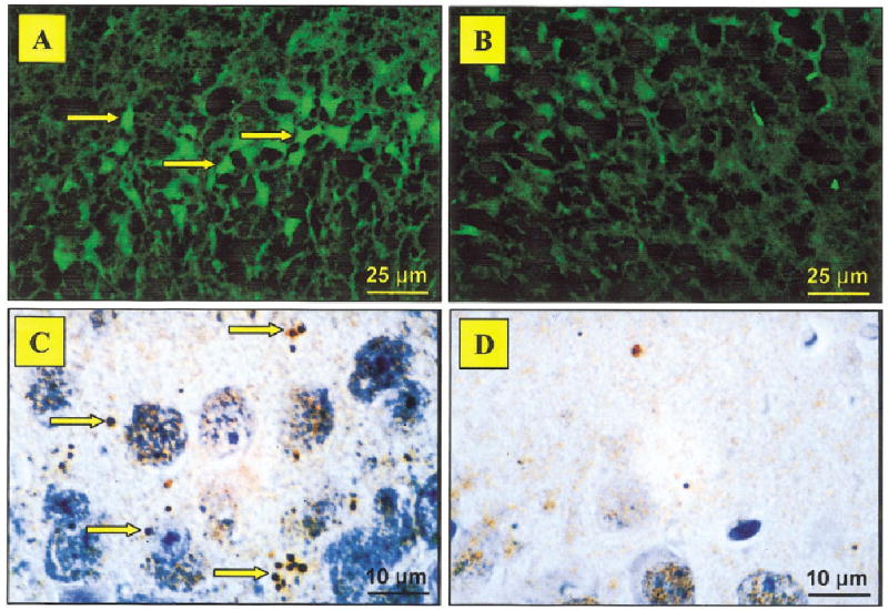
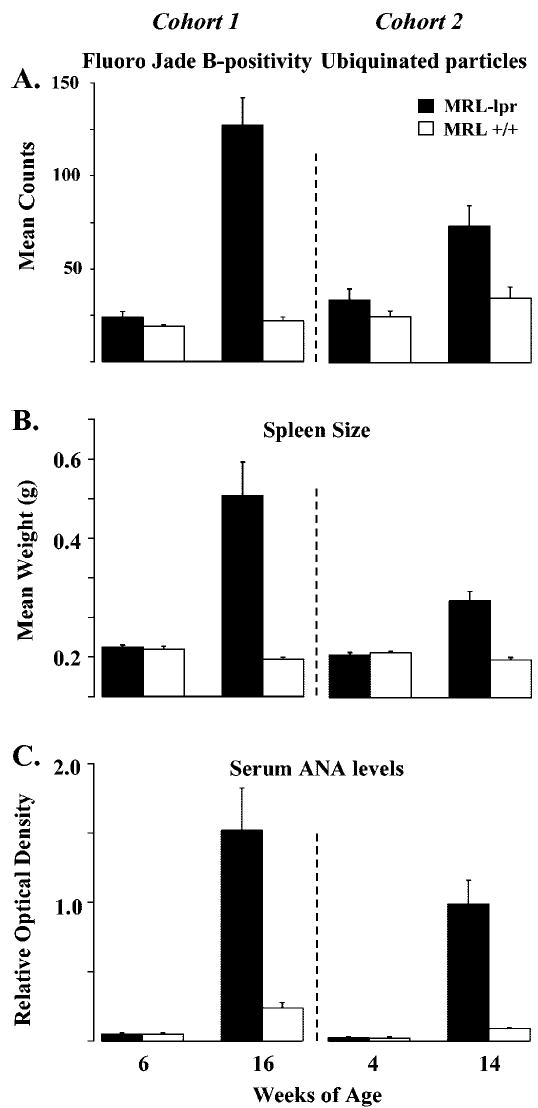
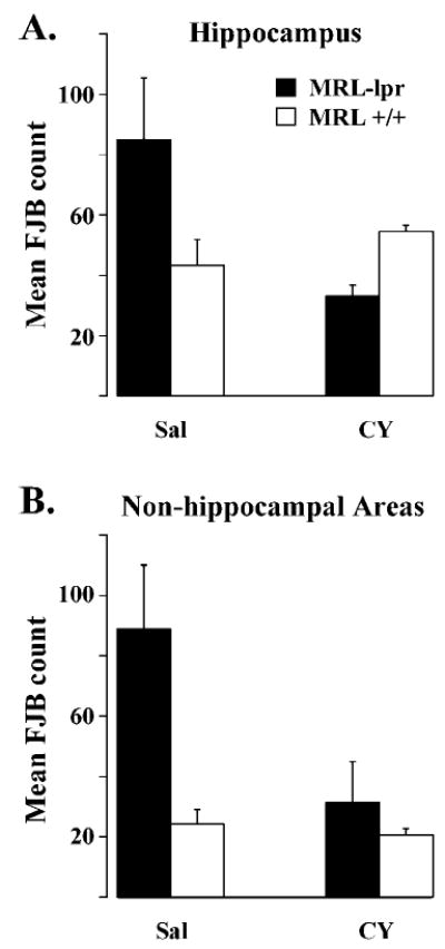
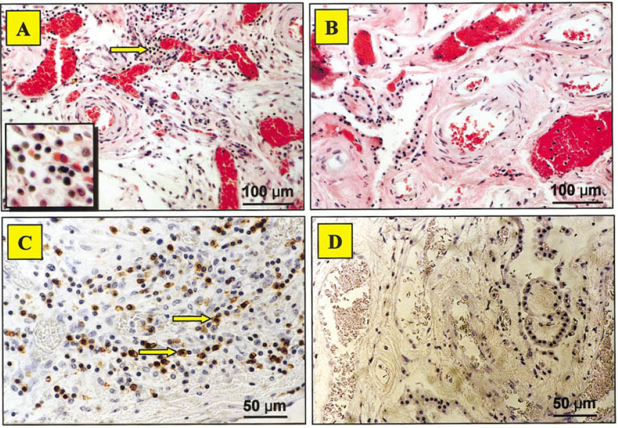
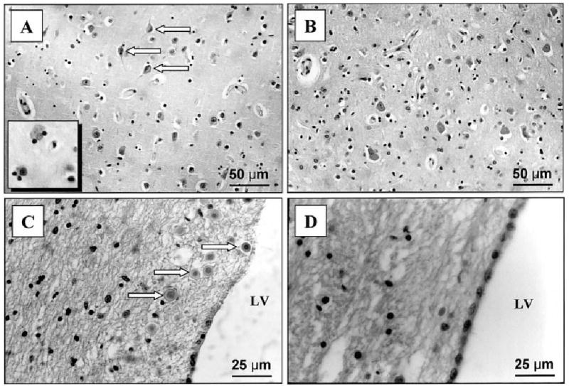
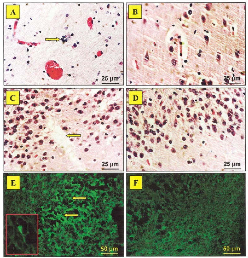
Systemic lupus erythematosus (SLE) is frequently accompanied by neuropsychiatric (NP) and cognitive deficits of unknown etiology. By using autoimmune MRL-lpr mice as an animal model of NP-SLE, we examine the relationship between autoimmunity, hippocampal damage, and behavioral dysfunction. Fluoro Jade B (FJB) staining and anti-ubiquitin (anti-Ub) immunocytochemistry were used to assess neuronal damage in young (asymptomatic) and aged (diseased) mice, while spontaneous alternation behavior (SAB) was used to estimate the severity of hippocampal dysfunction. The causal relationship between autoimmunity and neuropathology was tested by prolonged administration of the immunosuppressive drug cyclophosphamide (CY). In comparison to congenic MRL +/+ controls, SAB acquisition rates and performance in the "reversal" trial were impaired in diseased MRL-lpr mice, suggesting limited use of the spatial learning strategy. FJB-positive neurons and anti-Ub particles were frequent in the CA3 region. Conversely, CY treatment attenuated the SAB deficit and overall FJB staining. Similarly to mouse brain, the hippocampus from a patient who died from NP-SLE showed reduced neuronal density in the CA3 region and dentate gyrus, as well as increased FJB positivity in these regions. Gliosis and neuronal loss were observed in the gray matter, and T lymphocytes and stromal calcifications were common in the choroid plexus. Taken together, these results suggest that systemic autoimmunity induces significant hippocampal damage, which may underlie affective and cognitive deficits in NP-SLE.
Figures
Similar articles
-
Neurodegeneration in autoimmune MRL-lpr mice as revealed by Fluoro Jade B staining.Brain Res. 2003 Feb 28;964(2):200-10. doi: 10.1016/s0006-8993(02)03980-x. Brain Res. 2003. PMID: 12576180
-
Impaired response to amphetamine and neuronal degeneration in the nucleus accumbens of autoimmune MRL-lpr mice.Behav Brain Res. 2006 Jan 6;166(1):32-8. doi: 10.1016/j.bbr.2005.07.030. Epub 2005 Sep 23. Behav Brain Res. 2006. PMID: 16183144 Free PMC article.
-
Autoimmune-induced damage of the midbrain dopaminergic system in lupus-prone mice.J Neuroimmunol. 2004 Jul;152(1-2):83-97. doi: 10.1016/j.jneuroim.2004.04.003. J Neuroimmunol. 2004. PMID: 15223241
-
The MRL/lpr mouse strain as a model for neuropsychiatric systemic lupus erythematosus.J Biomed Biotechnol. 2011;2011:207504. doi: 10.1155/2011/207504. Epub 2011 Feb 10. J Biomed Biotechnol. 2011. PMID: 21331367 Free PMC article. Review.
-
Systemic lupus erythematosus involving the nervous system: presentation, pathogenesis, and management.Clin Rev Allergy Immunol. 2008 Jun;34(3):356-60. doi: 10.1007/s12016-007-8052-z. Clin Rev Allergy Immunol. 2008. PMID: 18181036 Review.
Cited by
-
From Systemic Inflammation to Neuroinflammation: The Case of Neurolupus.Int J Mol Sci. 2018 Nov 13;19(11):3588. doi: 10.3390/ijms19113588. Int J Mol Sci. 2018. PMID: 30428632 Free PMC article. Review.
-
C5a alters blood-brain barrier integrity in experimental lupus.FASEB J. 2010 Jun;24(6):1682-8. doi: 10.1096/fj.09-138834. Epub 2010 Jan 11. FASEB J. 2010. PMID: 20065106 Free PMC article.
-
Altered hippocampal connectivity dynamics predicts memory performance in neuropsychiatric lupus: a resting-state fMRI study using cross-recurrence quantification analysis.Lupus Sci Med. 2023 Jul;10(2):e000920. doi: 10.1136/lupus-2023-000920. Lupus Sci Med. 2023. PMID: 37400223 Free PMC article.
-
Sustained Immunosuppression Alters Olfactory Function in the MRL Model of CNS Lupus.J Neuroimmune Pharmacol. 2017 Sep;12(3):555-564. doi: 10.1007/s11481-017-9745-6. Epub 2017 Apr 11. J Neuroimmune Pharmacol. 2017. PMID: 28401431
-
Animal models of neuropsychiatric systemic lupus erythematosus: deciphering the complexity and guiding therapeutic development.Autoimmunity. 2024 Dec;57(1):2330387. doi: 10.1080/08916934.2024.2330387. Epub 2024 Mar 31. Autoimmunity. 2024. PMID: 38555866 Review.
References
-
- Alexander EL, Murphy ED, Roths JB, Alexander GE. Congenic autoimmune murine models of central nervous system disease in connective tissue disorders. Ann Neurol. 1983;14:242–248. - PubMed
-
- Alves-Rodrigues A, Gregori L, Figueiredo-Pereira ME. Ubiquitin, cellular inclusions and their role in neurodegeneration. Trends Neurosci. 1998;21:516–520. - PubMed
-
- Armanini MP, Hutchins C, Stein BA, Sapolsky RM. Glucocorticoid endangerment of hippocampal neurons is NMDA-receptor dependent. Brain Res. 1990;532:7–12. - PubMed
-
- Atkins CJ, Kondon JJ, Quismorio FP, Friou GJ. The choroid plexus in systemic lupus erythematosus. Ann Intern Med. 1972;76:65–72. - PubMed
Publication types
MeSH terms
Substances
Grants and funding
LinkOut - more resources
Full Text Sources
Medical
Miscellaneous
