. 2004 Aug;122(8):1190-200.
doi: 10.1001/archopht.122.8.1190.

Retinal degeneration in a rodent model of Smith-Lemli-Opitz syndrome: electrophysiologic, biochemical, and morphologic features

Affiliations

Retinal degeneration in a rodent model of Smith-Lemli-Opitz syndrome: electrophysiologic, biochemical, and morphologic features

Steven J Fliesler et al. Arch Ophthalmol. 2004 Aug.

Abstract

Objective: To assess the electrophysiologic, histologic, and biochemical features of an animal model of Smith-Lemli-Opitz syndrome (SLOS).

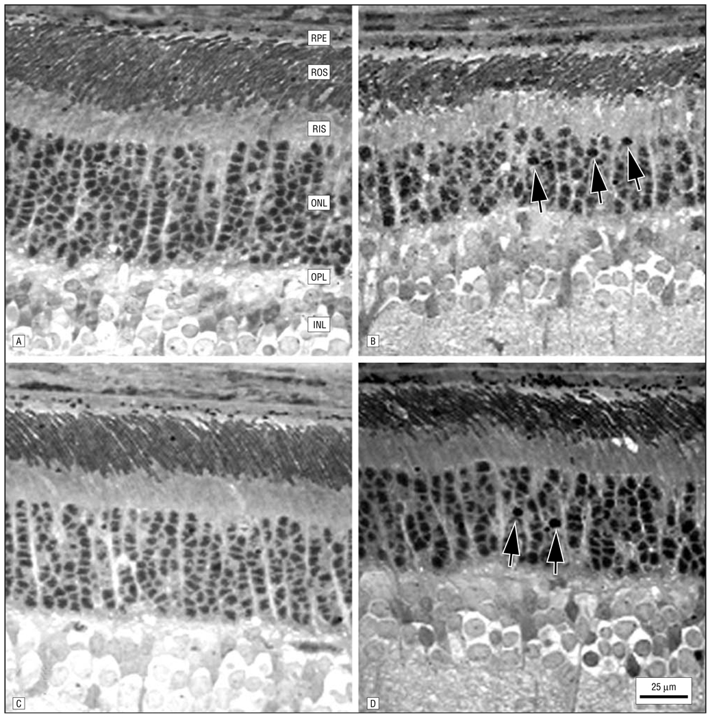
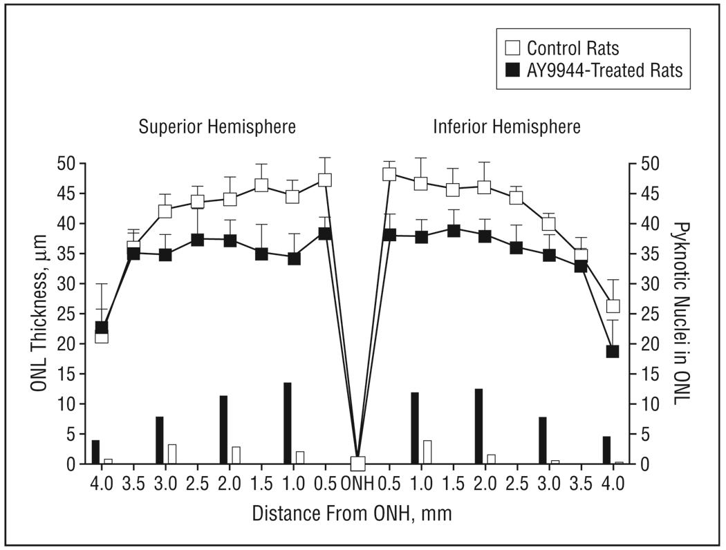
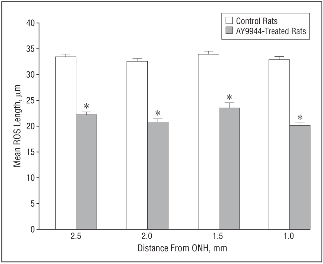
Methods: Sprague-Dawley rats were treated with AY9944, a selective inhibitor of 3beta-hydroxysterol-Delta(7)-reductase (the affected enzyme in SLOS). Dark- and light-adapted electroretinograms were obtained from treated and control animals. From each animal, 1 retina was analyzed by microscopy, and the contralateral retina plus serum samples were analyzed for sterol composition. The main outcome measures were rod and cone electroretinographic amplitudes and implicit times, outer nuclear layer (ONL) thickness, rod outer segment length, pyknotic ONL nucleus counts, and the 7-dehydrocholesterol/cholesterol mole ratio in the retina and serum.

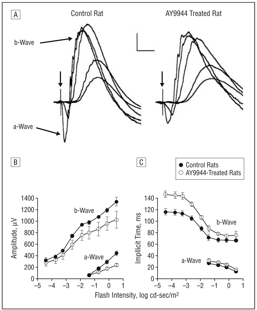
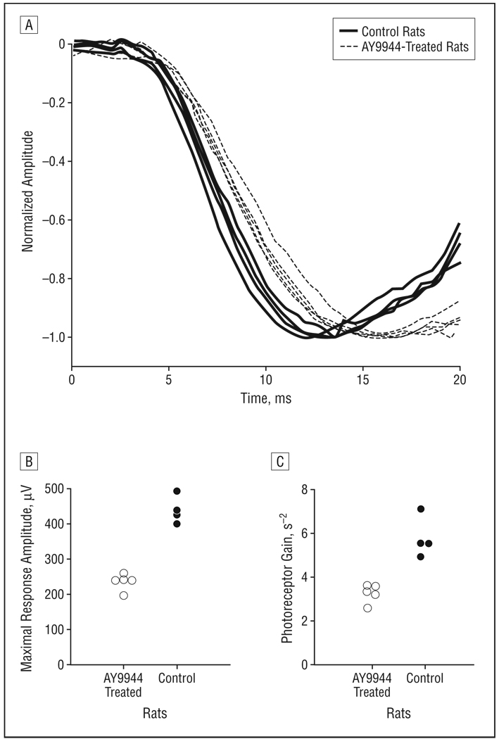
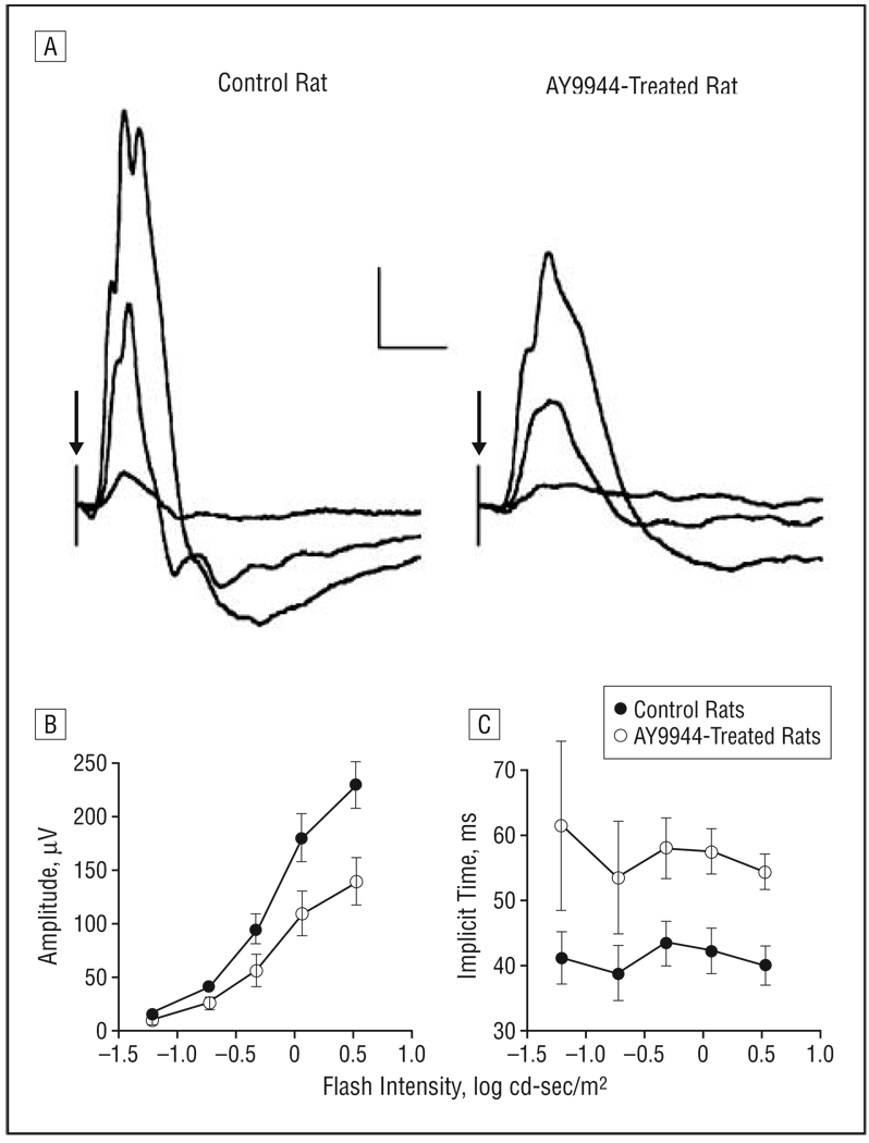
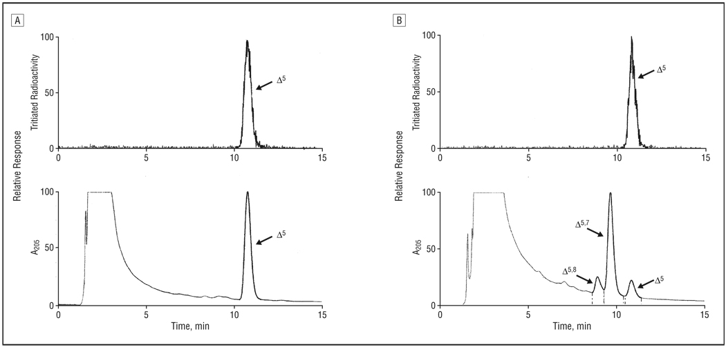
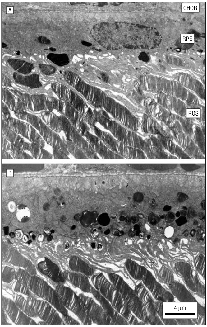
Results: By 10 weeks' postnatal age, rod and cone electroretinographic wave amplitudes in AY9944-treated animals were significantly reduced and implicit times were significantly increased relative to controls. Maximal rod photoresponse and gain values were reduced approximately 2-fold in treated animals relative to controls. The ONL thickness and average rod outer segment length were reduced by approximately 18% and 33%, respectively, and ONL pyknotic nucleus counts were approximately 4.5-fold greater in treated animals relative to controls. The retinal pigment epithelium of treated animals contained massive amounts of membranous/lipid inclusions not routinely observed in controls. The 7-dehydrocholesterol/cholesterol mole ratios in treated retinas and serum samples were approximately 5:1 and 9:1, respectively, whereas the ratios in control tissues were essentially zero.

Conclusions: This rodent model exhibits the key biochemical hallmarks associated with SLOS and displays electrophysiologic deficits comparable to or greater than those observed in the human disease. Clinical Relevance These results predict retinal degeneration in patients with SLOS, particularly those with the more severe (type II) form of the disease, and may be more broadly relevant to other inborn errors of cholesterol biosynthesis. This animal model may also be of use in evaluating therapeutic treatments for SLOS and in understanding the slow phototransduction kinetics observed in patients with SLOS.


Figures

Figure 1
Figure 1
Dark-adapted electroretinograms. A, Representative electroretinograms recorded from a control rat and a rat treated with AY9944 (trans-1,4-bis [2-dichlorobenzylamino-ethyl] cyclohexane dihydrochloride) to strobe flashes presented to the dark-adapted eye. Calibration indicates 200 µV and 50 milliseconds. In the flash intensity–response functions for a- and b-wave amplitudes (B) and implicit time (C), each point represents the mean value for 4 control rats and 5 rats treated with AY9944. Error bars represent SD; cd, candela.
Figure 2
Figure 2
Analysis of the leading edge of the rod electroretinographic a-wave. A, Initial portion of the rod electroretinogram obtained to a 0.5–log candela (cd)-sec/m2 strobe flash, the highest flash intensity used in this study. Each waveform was obtained from a different rat, and each has been normalized by the maximal response amplitude. Distribution of values of is shown for maximal response amplitude (B) and photoreceptor gain (C) obtained for control rats and rats treated with AY9944 (trans-1,4-bis [2-dichlorobenzylamino-ethyl] cyclohexane dihydrochloride).
Figure 3
Figure 3
Light-adapted electroretinograms. A, Representative cone electroretinograms recorded from a control rat and a rat treated with AY9944 (trans-1,4-bis [2-dichlorobenzylamino-ethyl] cyclohexane dihydrochloride) to strobe flashes superimposed on a steady rod-desensitizing adapting field. Calibration indicates 50 µV and 50 milliseconds. In flash intensity–response functions for cone amplitude (B) and implicit time (C), each point represents the mean value for 4 control rats and 5 rats treated with AY9944. Error bars represent SD; cd, candela.
Figure 4
Figure 4
Reverse-phase high-performance liquid chromatograms of nonsaponifiable lipid extracts from retinas of control rats (A) and AY9944 (trans-1,4-bis [2-dichlorobenzylamino-ethyl] cyclohexane dihydrochloride)-treated rats (B). The upper panels show the radioactivity detector response, demonstrating chromatographic elution of the [3H] cholesterol internal standard; the lower panels, the UV detector response (absorbance at 205 nm). Elution positions of cholesterol (Δ5), 7-dehydrocholesterol (Δ5, 7), and 8-dehydrocholesterol (Δ5, 8) are indicated. Full-scale detector response is set for the maximum response of the dominant sterol component in each panel.
Figure 5
Figure 5
Histologic changes in retinas from 3-month-old control (A and C) and AY9944 (trans-1,4-bis [2-dichlorobenzylamino-ethyl] cyclohexane dihydrochloride)-treated (B and D) rats corresponding to regions 2 mm from the optic nerve head in the superior (A and B) and inferior (C and D) hemispheres along the vertical meridian. Note the apparent reduction in outer nuclear layer (ONL) thickness and rod outer segment (ROS) length in the retinal regions of the AY9944-treated rat relative to the comparable regions of the control retina and the presence of pyknotic nuclei (arrows) in retinas of treated rats. RPE indicates retinal pigment epithelium; RIS, rod inner segment layer; OPL, outer plexiform layer; and INL, inner nuclear layer.
Figure 6
Figure 6
Morphometric analysis of the outer nuclear layer (ONL) of retinas from control and AY9944 (trans-1,4-bis [2-dichlorobenzylamino-ethyl] cyclohexane dihydrochloride)-treated rats (aged 3 months). Mean ONL thickness values (plotted curves, 15 independent measurements per region) are measured at 0.5-mm intervals from the optic nerve head (ONH) along the vertical meridian. Mean pyknotic ONL nucleus counts (bar graphs) are given per 1-mm expanse along the vertical meridian. Error bars represent SD.
Figure 7
Figure 7
Morphometric analysis of mean rod outer segment (ROS) length in retinas from control and AY9944 (trans-1,4-bis [2-dichlorobenzylaminoethyl] cyclohexane dihydrochloride)-treated rats. Measurements (N=30) were taken in the superior retinal hemisphere at 0.5-mm intervals along the vertical meridian, 1.0 to 2.5 mm from the optic nerve head (ONH). Asterisk ndicates a statistically significant difference in ROS length measurements in control vs treated retinas at each retinal region examined (P<.001). Error bars represent SD.
Figure 8
Figure 8
Ultrastructure of the retina and underlying retinal pigment epithelium (RPE)–choroid (CHOR) in a 3-month-old control rat (A) and an age-matched AY9944 (trans-1,4-bis [2-dichlorobenzylamino-ethyl] cyclohexane dihydrochloride)-treated rat (B). Note the marked accumulation of membranous and lipid inclusions in the RPE of the treated rat eye. ROS indicates rod outer segment.

Similar articles

Cited by

References

    1. Smith JD, Lemli L, Opitz JM. A newly recognized syndrome of multiple congenital anomalies. J Pediatr. 1964;64:210–217. - PubMed
    1. Kelley RI. Inborn errors of cholesterol biosynthesis. Adv Pediatr. 2000;47:1–53. - PubMed
    1. Porter FD. Human malformation syndromes due to inborn errors of cholesterol synthesis. Curr Opin Pediatr. 2003;15:607–613. - PubMed
    1. Herman GE. Disorders of cholesterol biosynthesis: prototypic metabolic malformation syndromes. Hum Mol Genet. 2003;12 suppl 1:R75–R88. - PubMed
    1. Battaile KP, Battaile BC, Merkens LS, Maslen CL, Steiner RD. Carrier frequency of the common mutation IVS8-1G>C in DHCR7 and estimate of the expected incidence of Smith-Lemli-Opitz syndrome. Mol Genet Metab. 2001;72:67–71. - PubMed

Publication types

MeSH terms

Substances

Associated data