. 2004 Aug;3(4):855-61.
doi: 10.1128/EC.3.4.855-861.2004.

Multiple triclosan targets in Trypanosoma brucei

Affiliations

Multiple triclosan targets in Trypanosoma brucei

Kimberly S Paul et al. Eukaryot Cell. 2004 Aug.

Abstract

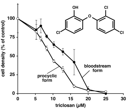
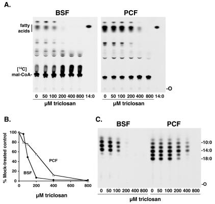
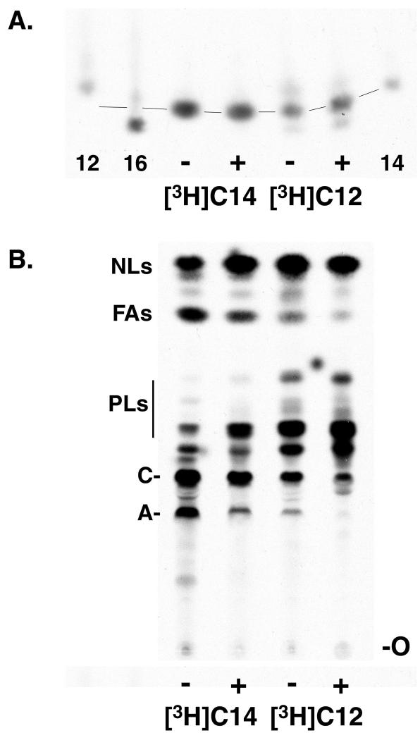
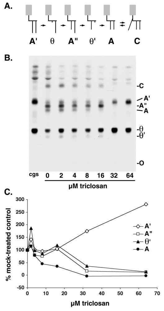
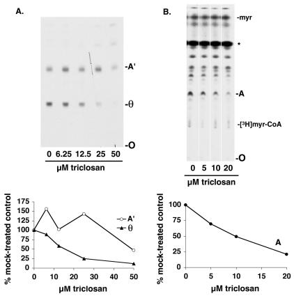
Trypanosoma brucei genes encoding putative fatty acid synthesis enzymes are homologous to those encoding type II enzymes found in bacteria and organelles such as chloroplasts and mitochondria. It was therefore not surprising that triclosan, an inhibitor of type II enoyl-acyl carrier protein (enoyl-ACP) reductase, killed both procyclic forms and bloodstream forms of T. brucei in culture with 50% effective concentrations (EC(50)s) of 10 and 13 microM, respectively. Triclosan also inhibited cell-free fatty acid synthesis, though much higher concentrations were required (EC(50)s of 100 to 200 microM). Unexpectedly, 100 microM triclosan did not affect the elongation of [(3)H]laurate (C(12:0)) to myristate (C(14:0)) in cultured bloodstream form parasites, suggesting that triclosan killing of trypanosomes may not be through specific inhibition of enoyl-ACP reductase but through some other mechanism. Interestingly, 100 microM triclosan did reduce the level of incorporation of [(3)H]myristate into glycosyl phosphatidylinositol species (GPIs). Furthermore, we found that triclosan inhibited fatty acid remodeling in a cell-free assay in the same concentration range required for killing T. brucei in culture. In addition, we found that a similar concentration of triclosan also inhibited the myristate exchange pathway, which resides in a distinct subcellular compartment. However, GPI myristoylation and myristate exchange are specific to the bloodstream form parasite, yet triclosan kills both the bloodstream and procyclic forms. Therefore, triclosan killing may be due to a nonspecific perturbation of subcellular membrane structure leading to dysfunction in sensitive membrane-resident biochemical pathways.


Figures

FIG. 1.
FIG. 1.
Inhibition of T. brucei growth in culture by triclosan. Cultures of T. brucei procyclic and bloodstream form trypanosomes were treated in duplicate for 48 h with either triclosan or NaOH as a solvent control, and cell density was assessed by hemocytometer counting. Cell densities are expressed as percentages of the density of mock-treated culture. The inset shows the structure of triclosan.
FIG. 2.
FIG. 2.
Inhibition of cell-free fatty acid synthesis by triclosan. Bloodstream form (BSF) and procyclic form (PCF) membranes were preincubated with various concentrations of triclosan and assayed for fatty acid synthesis. (A) Lipids were extracted and analyzed by TLC. 14:0, [3H]myristate marker; O, origin; Mal-CoA, malonyl-CoA. (B) Densitometric quantitation of fatty acids from panel A. (C) Chain length analysis of the fatty acids shown in panel A. Fatty acid methyl esters were prepared and analyzed by reverse-phase TLC. The labels 10:0, 14:0, and 18:0 indicate the positions of [methyl-3H]decanoate, [methyl-3H]myristate, and [methyl-3H]stearate, respectively.
FIG. 3.
FIG. 3.
Inhibition of metabolic labeling of glycolipids A and C. (A) Cultured bloodstream trypanosomes were metabolically labeled in the absence (−) or presence (+) of 100 μM triclosan with [3H]myristate ([3H]C14), or [3H]laurate ([3H]C12). Lipids were extracted, fatty acid methyl esters were prepared, and fatty acid chain lengths were analyzed by reverse-phase TLC. Only a portion of the TLC is shown; the rest of the TLC was blank. The labels 12, 14, and 16 indicate [methyl-3H]laurate, [methyl-3H]myristate, and [methyl-3H]palmitate, respectively. Lines indicate the pattern of “smiling” on the TLC. (B) Total lipid extracts from the same cells analyzed for panel A were analyzed by normal-phase TLC. NLs, neutral lipids; FAs, free fatty acids; PLs, phospholipids; C, glycolipid C; A, glycolipid A; O, origin.
FIG. 4.
FIG. 4.
Inhibition of cell-free fatty acid remodeling by triclosan. (A) Scheme of fatty acid remodeling pathway of the VSG GPI anchor. The dimyristoylated GPI anchor precursor glycolipid A is generated from glycolipid A′ (whose fatty acids are longer than myristate) through two rounds of deacylation and myristoylation. See reference for an alternative remodeling pathway involving inositol-acylated GPIs. Gray boxes represent the glycan portion of the GPI, short black vertical lines represent myristate, and long black vertical lines represent fatty acids longer than myristate. (B) Bloodstream form membranes were incubated with GDP-[3H]mannose to generate radiolabeled glycolipids A′ and θ, the substrates for fatty acid remodeling (see core glycan synthesis [cgs] lane). The lysates were then incubated for 30 min on ice with various amounts of triclosan prior to the addition of myristoyl-CoA. Glycolipids were extracted and analyzed by normal-phase TLC. The faster-migrating bands above glycolipid A′ are intermediates in core glycan synthesis and inositol-acylated species. O, origin. (C) Quantitation of the results of TLC shown in panel B, obtained by densitometry. Values are expressed as a percentage of the value obtained with a 0 μM triclosan control reaction.
FIG. 5.
FIG. 5.
Inhibition of θ formation and myristate exchange by triclosan. (A, upper panel) Bloodstream form membranes were preincubated with various amounts of triclosan and then incubated with GDP-[3H]mannose to generate radiolabeled glycolipids A′ and θ, the substrates for fatty acid remodeling. Glycolipids were extracted and analyzed by normal-phase TLC. (Lower panel) Quantitation by densitometry of the results of TLC shown in the upper panel. Values are expressed as a percentage of the value obtained with a 0 μM triclosan control reaction. A′ and θ, glycolipids A′ and θ; O, origin. (B, upper panel) Bloodstream form membranes were preincubated with various amounts of triclosan and assayed for myristate exchange by the addition of [3H]myristate, ATP, and CoA. Glycolipids were extracted and analyzed by normal-phase TLC. The species marked with an asterisk (*) is formed by boiling the samples (see reference 34). myr, myristate; A, glycolipid A; O, origin. The other unidentified species labeled with [3H]myristate are composed of neutral lipids, phospholipids, and other glycolipid species. (Lower panel) Quantitation by densitometry of the results of TLC shown in the upper panel. Values are expressed as a percentage of the value obtained with a 0 μM triclosan control reaction.

References

    1. Bhargava, H. N., and P. A. Leonard. 1996. Triclosan: applications and safety. Am. J. Infect. Control 24:209-218. - PubMed
    1. Brun, R., and M. Shonenberger. 1979. Cultivation and in vitro cloning of procyclic culture forms of Trypanosoma brucei in a semi-defined medium. Acta Trop. 36:289-292. - PubMed
    1. Buxbaum, L. U., K. G. Milne, K. A. Werbovetz, and P. T. Englund. 1996. Myristate exchange on the Trypanosoma brucei variant surface glycoprotein. Proc. Natl. Acad. Sci. USA 93:1178-1183. - PMC - PubMed
    1. Buxbaum, L. U., J. Raper, F. R. Opperdoes, and P. T. Englund. 1994. Myristate exchange: a second glycosyl phosphatidylinositol myristoylation reaction in African trypanosomes. J. Biol. Chem. 269:30212-30220. - PubMed
    1. Doering, T. L., M. S. Pessin, G. W. Hart, D. M. Raben, and P. T. Englund. 1994. The fatty acids in unremodeled trypanosome glycosyl phosphatidylinositols. Biochem. J. 299:741-746. - PMC - PubMed

Publication types

MeSH terms

LinkOut - more resources