Comparative Study
. 2004 Aug 16;200(4):459-67.
doi: 10.1084/jem.20040342. Epub 2004 Aug 9.

Masking of phosphatidylserine inhibits apoptotic cell engulfment and induces autoantibody production in mice

Affiliations
Comparative Study

Masking of phosphatidylserine inhibits apoptotic cell engulfment and induces autoantibody production in mice

Kenichi Asano et al. J Exp Med. .

Abstract

Apoptotic cells are rapidly phagocytosed by professional phagocytes, such as macrophages and dendritic cells. This process prevents the release of potentially noxious or immunogenic intracellular materials from dying cells, and is thought to play a critical role for the maintenance of normal functions in surrounding tissues. Milk fat globule-EGF-factor 8 (MFG-E8), secreted by activated macrophages and immature dendritic cells, links apoptotic cells and phagocytes, and promotes phagocytosis of apoptotic cells. Here, we report that an MFG-E8 mutant, designated as D89E, carrying a point mutation in an RGD motif, inhibited not only the phagocytosis of apoptotic cells by a wide variety of phagocytes, but also inhibited the enhanced production of IL-10 by thioglycollate-elicited peritoneal macrophages phagocytosing apoptotic cells. When intravenously injected into mice, the D89E protein induced the production of autoantibodies including antiphospholipids antibodies and antinuclear antibodies. The production of autoantibodies was enhanced by the coinjection of syngeneic apoptotic thymocytes. After the induction of autoantibody production by D89E, the treated mice showed a long-term elevation of the titer for autoantibodies, and developed IgG deposition in the glomeruli. These results indicated that the impairment of apoptotic cell phagocytosis led to autoantibody production.


Figures
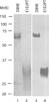
Figure 1.
Figure 1.
Preparation of recombinant MFG-E8. The expression plasmids of D89E carrying the RGE sequence instead of RGD in the second EGF-like domain of mouse MFG-E8, or E1E2PT lacking the C1C2 domains were introduced into human 293T cells. Recombinant proteins in the supernatants were purified using anti-FLAG M2 affinity gel. D89E (lanes 1 and 3) or E1E2PT (lanes 2 and 4) were separated by SDS-PAGE on a 10/20% polyacrylamide gradient gel and stained with CBB (lanes 1 and 2), or transferred to a PVDF membrane and detected with anti-FLAG antibody (lanes 3 and 4). Positions of molecular mass standard proteins are indicated in kilodaltons on the left.
Figure 2.
Figure 2.
The D89E inhibits phagocytosis of apoptotic cells by macrophages. (A) Mouse bone marrow–derived macrophages (BMDM) or (B) resident peritoneal macrophages (Resident pMac) were pretreated for 30 min with indicated concentrations of D89E or E1E2PT and were cocultured with dexamethasone-treated apoptotic thymocytes from CAD-deficient mice. The cells were stained for Mac-1 and TUNEL, analyzed by flow cytometry, and the percentage of TUNEL-positive cells in the Mac-1–positive population was determined. The assay was performed in triplicate, and mean values are shown with SD. These results are representative of three independent experiments.
Figure 3.
Figure 3.
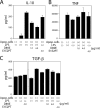
D89E inhibits the enhanced production of IL-10 in macrophages that engulf apoptotic cells. Thioglycollate-elicited mouse peritoneal macrophages were cocultured with or without UV-irradiated thymocytes for 2 h in the absence or presence of indicated concentrations of the D89E or E1E2PT protein. After washing twice, the macrophages were treated with or without 1 μg/ml of LPS for 20 h. The concentrations of IL-10 (A), TNFα (B), and TGF-β (C) were determined by ELISA. The assay was performed in triplicate, and mean values are shown with SD. These results are representative of three independent experiments.
Figure 4.
Figure 4.
Intravenous injection of D89E induces autoantibody production in mice. Either D89E or E1E2PT (0.6 μg) were diluted in 300 μl PBS containing 2.5% normal syngeneic mouse serum (concentration, 2.0 μg /ml), and injected into C57BL/6 mice weekly for the total of four (A and B) or six (C–E) injections. Blood samples were obtained 1 wk after the last injection, and serum levels of anti-CL (A), anti-PS (B), and anti-dsDNA (E) were evaluated by ELISA as described in Materials and Methods. ANAs were detected by indirect immunofluorescence on Hep-2 cells (C) and ELISA (D). Serum levels of autoantibodies in MRL/Mpj-lpr/lpr mice (16–40 wk old) are shown as a control. Horizontal bars represent the means. Statistical significance was determined by a one-tailed Mann-Whitney test. P indicates a difference with a P < 0.01.
Figure 5.
Figure 5.
Dose-dependent effects of D89E on the development of autoantibodies. The D89E protein was diluted in PBS containing 2.5% normal syngeneic mouse serum at a concentration of 0.125, 0.5, or 2.0 μg/ml, and 300 μl of the solution was injected intravenously into C57BL/6 mice. Mice were given six injections at weekly intervals and bled a week after the last immunization. Serum levels of anti-CL (A) and ANA (B) were measured by ELISA. *P indicates a difference with a P < 0.05.
Figure 6.
Figure 6.
Coinjection of apoptotic cells augments autoantibody production induced by D89E. Thymocytes were prepared from C57BL/6 mice, cultured in RPMI 1640 containing 1% syngeneic mouse serum, and irradiated with 40 J/m2 UV light. After incubation for 20 h to induce apoptosis, the indicated number of cells was intravenously injected with or without 0.6 μg of D89E protein into syngeneic mice. Serum levels of anti-CL (A) and ANA (B) were measured by ELISA after four or six weekly injections, respectively. Horizontal bars represent the means. *P indicates a difference with a P < 0.05.
Figure 7.
Figure 7.
Sustained elevation of autoantibodies in mice injected with D89E. Titers of autoantibodies were followed over 32 wk after immunization. C57BL/6 mice were injected intravenously with 300 μl of 2.0 μg/ml D89E. Injections were administered weekly for a total of four immunizations (arrows). Mice were bled 2, 4, 9, 18, 25, 30, and 32 wk after the first immunization. Anti-CL (A) and ANA (B) were measured by ELISA as described in Materials and Methods.
Figure 8.
Figure 8.
Immunohistochemical detection of IgG deposition in the glomeruli of kidneys in mice injected with D89E. C57BL/6 mice were injected intravenously with D89E or E1E2PT with or without apoptotic thymocytes six times at weekly intervals. Kidneys obtained 17 wk after the initial injection were fixed with 4% paraformaldehyde and embedded in paraffin. The sections were stained with Cy3-conjugated F(ab′)2 of goat anti-mouse IgG and were observed with fluorescence microscopy. 25 wk-old MRL/lpr mice (E) were used as a positive control. (A) 0.5 μg/ml D89E, (B) 2.0 μg/ml E1E2PT, (C) 2.0 × 10⁷ UV-irradiated thymocytes, and (D) 2.0 μg/ml D89E with 2.0 × 10⁷ of UV-irradiated thymocytes.

References

    1. Jacobson, M.D., M. Weil, and M.C. Raff. 1997. Programmed cell death in animal development. Cell. 88:347–354. - PubMed
    1. Vaux, D.L., and S.J. Korsmeyer. 1999. Cell death in development. Cell. 96:245–254. - PubMed
    1. Stroh, C., and K. Schulze-Osthoff. 1998. Death by a thousand cuts: an ever increasing list of caspase substrates. Cell Death Differ. 5:997–1000. - PubMed
    1. Nagata, S. 1997. Apoptosis by death factor. Cell. 88:355–365. - PubMed
    1. McIlroy, D., M. Tanaka, H. Sakahira, H. Fukuyama, M. Suzuki, K. Yamamura, Y. Ohsawa, Y. Uchiyama, and S. Nagata. 2000. An auxiliary mode of apoptotic DNA fragmentation provided by phagocytes. Genes Dev. 14:549–558. - PMC - PubMed

Publication types

MeSH terms