Comparative Study
. 2004 Aug 17;101(33):12236-41.
doi: 10.1073/pnas.0404998101. Epub 2004 Aug 9.

Evolution of p53 in hypoxia-stressed Spalax mimics human tumor mutation

Affiliations
Comparative Study

Evolution of p53 in hypoxia-stressed Spalax mimics human tumor mutation

Osnat Ashur-Fabian et al. Proc Natl Acad Sci U S A. .

Abstract

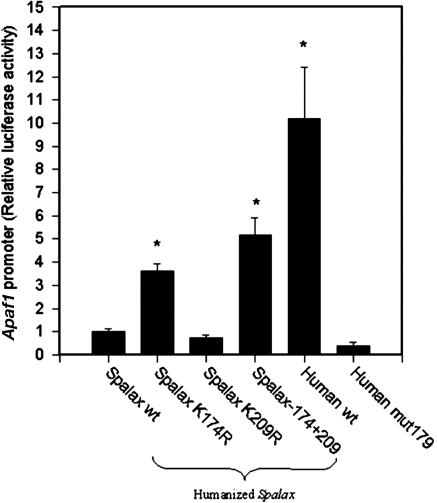
The tumor suppressor gene p53 controls cellular response to a variety of stress conditions, including DNA damage and hypoxia, leading to growth arrest and/or apoptosis. Inactivation of p53, found in 40-50% of human cancers, confers selective advantage under hypoxic microenvironment during tumor progression. The mole rat, Spalax, spends its entire life cycle underground at decidedly lower oxygen tensions than any other mammal studied. Because a wide range of respiratory adaptations to hypoxic stress evolved in Spalax, we speculated that it might also have developed hypoxia adaptation mechanisms analogous to the genetic/epigenetic alterations acquired during tumor progression. Comparing Spalax with human and mouse p53 revealed an arginine (R) to lysine (K) substitution in Spalax (Arg-174 in human) in the DNA-binding domain, identical to known tumor associated mutations. Multiple p53 sequence alignments with 41 additional species confirmed that Arg-174 is highly conserved. Reporter assays uncovered that Spalax p53 protein is unable to induce apoptosis-regulating target genes, resulting in no expression of apaf1 and partial expression of puma, pten, and noxa. However, cell cycle arrest and p53 stabilization/homeostasis genes were overactivated by Spalax p53. Lys-174 was found critical for apaf1 expression inactivation. A DNA-free p53 structure model predicts that Arg-174 is important for dimerization, whereas Spalax Lys-174 prevents such interactions. Similar neighboring mutations found in human tumors favor growth arrest rather than apoptosis. We hypothesize that, in an analogy with human tumor progression, Spalax underwent remarkable adaptive p53 evolution during 40 million years of underground hypoxic life.


Figures

Fig. 1.
Fig. 1.
Sequence of Spalax p53. Amino acid sequence alignment of Spalax (sp53), mouse (mp53), and human (hp53), with full-length p53 proteins (http://prodes.toulouse.inra.fr/multalin/multalin.html; ref. 25). The DNA-binding domain is shown in a box. Identities of species are in a white background. Changes among the species are in a gray background. Spalax-specific changes are depicted in bold black letters, and the amino acids common to other species are in bold white letters in a black background.
Fig. 2.
Fig. 2.
Relative transactivation of eight different human promoters. Human wild-type p53, mutated p53 (mut179 and mut342), and Spalax p53 were examined relative to an array of p53 target gene promoters. The relative transactivation with respect to human wild-type p53 is presented in a form similar to that of expression microarrays, ranging from complete loss of function (white) to a 6-fold increase of activation (red). Human wild-type p53 presents 1-fold of activation (blue), and other levels of transactivation are presented as shaded colors ranging from white (loss of function) to red (gain of function). Results are mean ± SE of four to eight independent transient transfections performed in triplicate (P value, unpaired t test).
Fig. 3.
Fig. 3.
Spalax wild-type and humanized p53 ability to transactivate Apaf1 promoter, with respect to human wild-type and mutated p53. Results are normalized to Renilla-luciferase and presented as fold-change from Spalax wild type (depicted as 1-fold of activation). Results are mean ± SE of four independent transfection assays, performed in triplicate *, P < 0.005 (unpaired t test).
Fig. 4.
Fig. 4.
Three-dimensional models of human, mouse, and Spalax proteins. (A) DNA-bound human p53 model. Arg-174 and Arg-209 are not located in or near the DNA-binding site and, thus, have no effect on the protein fold and/or stabilization in the DNA bound state. (BE) Protein–protein interaction between p53 DNA-binding domain monomers. Residues 174 (171 in the mouse) and 209 (206 in the mouse) and residues contacting them on the adjacent monomers are represented as ball-and-stick structures, colored by atom type (carbon in black, oxygen in red, and nitrogen in blue). (B) The human p53 dimer is displayed as cyan and green ribbons. Arg-174 contacts Arg-181 on the adjacent monomer. (C) The mouse P53 dimer (Left) is displayed as light blue and green ribbons. Arg-171 contacts Arg-178 on the adjacent monomer and is in close proximity to Glu-177. The modeled Spalax p53 dimer (purple and gold ribbons, Right) is shown in the same orientation as the human (D) and mouse (E) P53 dimer. The smaller Lys-174 displays different interaction within the interface, leading to a changed dimer configuration; thus, the dimer shown in the figure could not be preserved. Lys-209 is not involved in contacting the adjacent monomer displaying fewer contacts with Gly-181/Gly-187 on the adjacent monomers in humans and mice, respectively, with only a minor affect on the interface.

References

    1. Klein, G. (2004) Cell Death Differ. 11, 13-17. - PubMed
    1. Vousden, K. H. & Lu, X. (2002) Nat. Rev. Cancer 2, 594-604. - PubMed
    1. Levine, A. J. (1997) Cell 88, 323-331. - PubMed
    1. Graeber, T. G., Osmanian, C., Jacks, T., Housman, D. E., Koch, C. J., Lowe, S. W. & Giaccia, A. J. (1996) Nature 379, 88-91. - PubMed
    1. Kinzler, K. W. & Vogelstein, B. (1996) Nature 379, 19-20. - PubMed

Publication types

MeSH terms

Associated data