. 2004 Aug 17;101(33):12212-7.
doi: 10.1073/pnas.0404631101. Epub 2004 Aug 9.

A vascular cell-restricted RhoGAP, p73RhoGAP, is a key regulator of angiogenesis

Affiliations

A vascular cell-restricted RhoGAP, p73RhoGAP, is a key regulator of angiogenesis

Zhi-Jian Su et al. Proc Natl Acad Sci U S A. .

Abstract

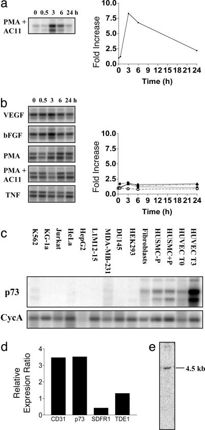
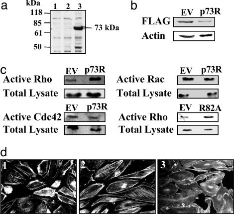
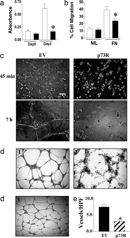
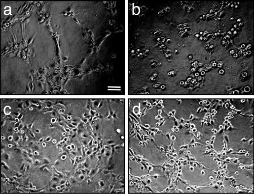
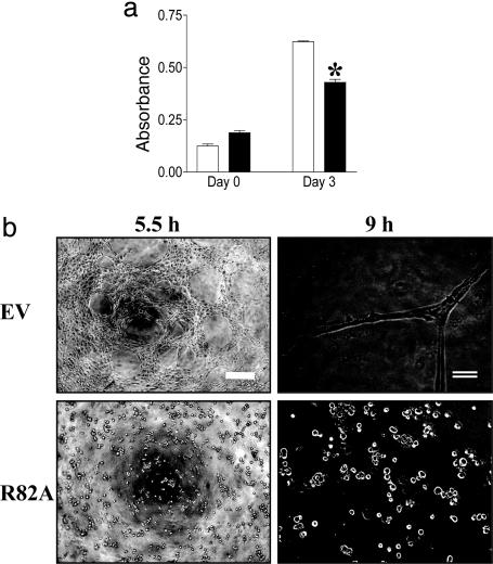
Angiogenesis is a major therapeutic target. Ideal drug targets are genes expressed only in endothelial cells (ECs) or only during the angiogenic process. Here, we describe a gene, p73RhoGAP (p73), that has both of these properties. By using a PCR-based subtraction-hybridization approach to clone cDNAs from ECs undergoing capillary-tube formation, we identified a RhoGAP member, p73. p73 displays GTPase activity to Rho but not to Rac or Cdc42. Knockdown of p73 protein, achieved by adenovirus delivery of p73 antisense and by small interfering RNA into ECs, demonstrated the importance of this protein in EC function. Under such conditions, EC migration, proliferation, and capillary-tube formation were inhibited. Furthermore, angiogenesis in vivo was also inhibited by antisense p73. A mutant R82A alteration achieved a similar phenotype in vitro to the antisense, demonstrating the importance of the GTPase-activating protein activity to p73 function. Expression profiling of p73 shows that it is vascular cell-selective, being highly expressed in ECs and smooth-muscle cells but not in other cell types. Finally, we show that the mRNA of p73 is up-regulated in an angiogenic milieu with little or no regulation seen under nonangiogenic conditions. p73, a vascular cell-specific GTPase-activating protein, is an important modulator of angiogenesis and displays many of features that make it worthy of being a drug target.


Figures

Fig. 1.
Fig. 1.
Expression of p73 during capillary-tube formation and its tissue distribution. (a and b) Virtual Northern blot analysis of p73 expression in capillary-tube formation. (a) HUVECs were plated onto a 3D collagen gel in the presence of PMA and anti-α2β1 to stimulate in vitro capillary-tube formation. Total RNA was harvested at the time points shown, and virtual Northern blots were probed with our isolated cDNA fragment. Blots were quantified by using a phosphorimager and standardized to cyclophilin A (CycA), and the fold increase was determined with respect to time given in the corresponding graph. (b) HUVECs, plated on a 2D collagen matrix, were treated with various stimuli, and the virtual Northern blots were probed as described for a. p73 levels were normalized to CycA, and the fold increase was plotted as follows: VEGF, ○; basic fibroblast growth factor (bFGF), ▪ ; PMA, ▴; PMA plus anti-α2β1 (PMA + AC11), ♦. TNF, tumor necrosis factor α. (c) Tissue distribution of p73 mRNA by virtual Northern blot analysis. Virtual Northern blots were generated from human cell lines and primary cells representing different cell types. HUVECs T0 and T3 refer to ECs at 0 and 3 h on 3D collagen gel. Human umbilical-vein smooth-muscle cells (HUSMCs) treated without and with PMA for 3 h are shown. (d) Expression of p73 in placenta, heart, lung, breast, colon, and HUVECs by using Q-RT-PCR. The ratio of expression in HUVECs over the median of the expression in the other tissues was calculated. This ratio was compared with the ratio obtained with the endothelial-selective marker CD31 (PECAM). (e) Northern blot analysis of p73 expression. Total HUVEC RNA (5 μg) was analyzed by Northern blot analysis by probing with the p73 3′ UTR-specific probe.
Fig. 2.
Fig. 2.
Characteristics and substrate specificity of p73. (a) p73 overexpression in 293 cells. EV and pAdtrack-Flag-p73 were transfected into 293 cells, and cell lysates were collected after 48 h. A Western blot was probed with anti-Flag M2 Ab (Sigma). Lanes: 1, cells alone; 2, EV; 3, Flag-p73. (b) p73 protein knockdown by p73R. EV and p73R were transfected into 293 cells and cotransfected with pAdtrack-Flag-p73 7 h later. Cell lysate was collected after 48 h. A Western blot was probed with anti-Flag M2 Ab, and anti-actin was used as a loading control. (c) p73 has activity for Rho but not Rac and Cdc42. HUVECs were adenovirally infected with EV, p73R, and p73 mutant (R82A), and they were then incubated without serum for 20 h. The cells were lysed and assayed for detection of active Rho-GTP, Rac-GTP, and Cdc42-GTP, and the average fold increase compared with EV is shown. Each result represents at least four independent experiments. (d) p73 regulates stress-fiber formation. HUVECs were adenovirally infected with EV (image 1) or p73R (images 2 and 3) 24 h before plating on a fibronectin. p73R-infected HUVECs were treated without (image 2) or with (image 3) C3 exoenzyme (30 μg/ml) for 32 h and stained with Rho–phalloidin.
Fig. 3.
Fig. 3.
p73 regulates angiogenesis. (a) p73R inhibits HUVEC proliferation. HUVECs were infected with EV (open bars) or p73R (filled bars) adenovirus, and proliferation assays were performed. Each bar is the mean ± SEM (n = 4) of three independent experiments. (b) p73R decreases HUVEC migration. HUVECs infected with EV (open bars) or p73R (filled bars) adenovirus were allowed to migrate toward either no chemotactic factor (NIL) or to fibronectin (FN). The number of migrated cells after 24 h was expressed as a percentage of the total cells added. Data are given as mean ± SEM (n = 3) of at least three independent experiments. (c) p73R inhibits tube formation. HUVECs infected with EV or p73R adenovirus were plated onto Matrigel, and capillary-tube formation was observed over a 12-h time course. Photographs were taken at 45 min and 7 h after plating. (Scale bar, 200 μm.) (d) Silencing of p73 by siRNA inhibits in vitro capillary-tube formation. HUVECs were transduced with either vector control (image 1), p73 siRNA (image 2), or control siRNA (image 3), selected for puromycin resistance, and after 4 days plated onto Matrigel. Tube formation after 24 h is shown and is representative of three experiments. (e) p73R inhibits blood-vessel invasion in vivo. Assessment of the number of blood vessels per high-power field (vessels/HPF) was made from three mice implanted with a VEGF with EV control adenovirus (black) and VEGF with p73R adenovirus (striped) impregnated gel-foam sponge. Results are given as mean ± SEM. Comparisons were made between each group and tested for significance by using a three-way nested ANOVA. *, P < 0.001.
Fig. 4.
Fig. 4.
The ROCK inhibitor, Y27632, reverses the p73 antisense phenotype. HUVECs were infected with either EV control (a and c) or p73 antisense (b and d) adenovirus. Cells were plated onto Matrigel in the absence (a and b) or presence (c and d) of the ROCK inhibitor Y27632 at a final concentration of 10μM. Formation of capillary tubes was assessed over the next 12 h. Photographs were taken at 4 h after plating.
Fig. 5.
Fig. 5.
p73 mutant inhibits EC function. (a) p73 (R82A) inhibits proliferation. EV-infected (open bars) and R82A-infected (filled bars) HUVECs were plated on day 0, and proliferation was measured 3 days later. Results are expressed as absorbance. Each bar represents the mean ± SEM (n = 4) of three independent experiments. (b) R82A inhibits tube formation. HUVECs infected with either EV or R82A were plated onto Matrigel, and capillary-tube formation was observed over 12 h. Photographs were taken at 5 and 9 h after plating. Scale bars are as follows: single line, 50 μm; double line, 200 μm. *, P < 0.05.

References

    1. Augustin, H. G. (1998) Trends Pharmacol. Sci. 19, 216-222. - PubMed
    1. Hanahan, D. (1997) Science 277, 48-50. - PubMed
    1. Gamble, J. R., Matthias, L. J., Meyer, G., Kaur, P., Russ, G., Faull, R., Berndt, M. C. & Vadas, M. A. (1993) J. Cell Biol. 121, 931-943. - PMC - PubMed
    1. Gamble, J., Meyer, G., Noack, L., Furze, J., Matthias, L., Kovach, N., Harlant, J. & Vadas, M. (1999) Endothelium 7, 23-34. - PubMed
    1. Bayless, K. J. & Davis, G. E. (2002) J. Cell Sci. 115, 1123-1136. - PubMed

Publication types

MeSH terms

LinkOut - more resources