. 2004 Aug 17;101(33):12130-5.
doi: 10.1073/pnas.0404720101. Epub 2004 Aug 9.

Large-scale characterization of HeLa cell nuclear phosphoproteins

Affiliations

Large-scale characterization of HeLa cell nuclear phosphoproteins

Sean A Beausoleil et al. Proc Natl Acad Sci U S A. .

Abstract

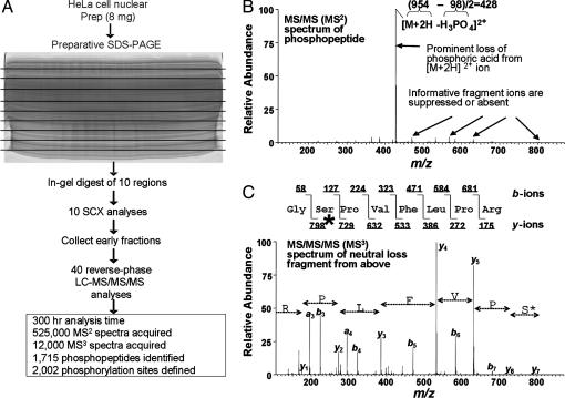
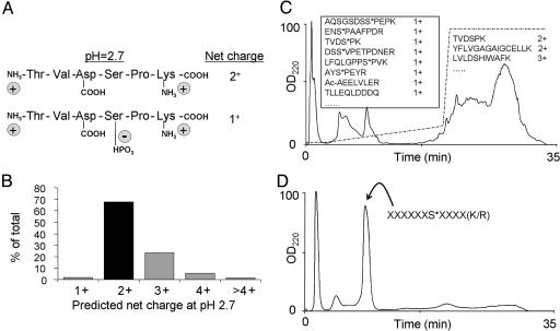
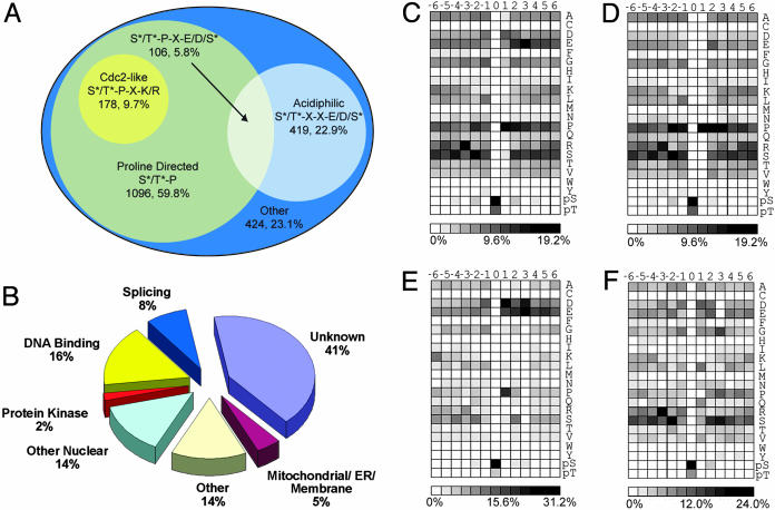
Determining the site of a regulatory phosphorylation event is often essential for elucidating specific kinase-substrate relationships, providing a handle for understanding essential signaling pathways and ultimately allowing insights into numerous disease pathologies. Despite intense research efforts to elucidate mechanisms of protein phosphorylation regulation, efficient, large-scale identification and characterization of phosphorylation sites remains an unsolved problem. In this report we describe an application of existing technology for the isolation and identification of phosphorylation sites. By using a strategy based on strong cation exchange chromatography, phosphopeptides were enriched from the nuclear fraction of HeLa cell lysate. From 967 proteins, 2,002 phosphorylation sites were determined by tandem MS. This unprecedented large collection of sites permitted a detailed accounting of known and unknown kinase motifs and substrates.


Figures

Fig. 2.
Fig. 2.
Analysis of human nuclear phosphorylation sites by multidimensional LC coupled to MS/MS/MS. (A) Eight milligrams of nuclear extract from asynchronous HeLa cells were separated by SDS/PAGE. The entire gel was excised into 10 regions and proteolyzed with trypsin followed by phosphopeptide enrichment by SCX LC. Early eluting fractions were subjected to amino acid sequence analysis by reverse-phase LC-MS/MS with data-dependent MS/MS/MS acquisition. Phosphorylation sites (n = 2,002) were identified by the sequest algorithm, acquisition of MS/MS/MS spectra, and manual validation. Prep, preparation. (B) Example of a MS/MS spectrum of a phosphopeptide showing a typical extensive neutral loss of phosphoric acid. (C)MS/MS/MS spectrum of the neutral loss precursor ion from B. Abundant peptide bond fragmentation permitted the unambiguous identification of this peptide from the protein cell division cycle 2-related protein kinase 7, with a phosphorylated Ser residue marked by an asterisk.
Fig. 1.
Fig. 1.
Scheme for phosphopeptide enrichment by SCX chromatography. (A) At pH 2.7, most peptides produced by trypsin proteolysis have a solution charge state of 2+, whereas phosphopeptides have a charge state of only 1+. (B) Solution charge state distribution of peptides (5–40 aa) produced by a theoretical digestion of the human protein database with trypsin (n = 6.8 × 10⁸ peptides). Sixty-eight percent of the predicted peptides have a net charge of 2+. Any peptide in this category would shift to a 1+ charge state upon phosphorylation. (C) SCX chromatography separation at pH 2.7 of a HeLa cell lysate after trypsin digestion. The dashed line indicates the salt gradient. Some identified peptides from the collected fractions are shown. Phosphorylation sites are denoted by an asterisk, and N-terminal acetylation is denoted by Ac. (D) SCX chromatography separation at pH 2.7 of a synthetic phosphopeptide library containing 2,000 phosphopeptides with a predicted solution charge state of 1+.
Fig. 3.
Fig. 3.
Classification of identified phosphorylation sites and amino acid frequencies surrounding phosphorylated Ser and Thr residues. From the 2,002 detected sites, 1,833 could be localized to a specific Ser or Thr without ambiguity. (A) Venn diagram representation of 1,833 precise sites of phosphorylation with respect to surrounding residues. Seventy-seven percent of the detected phosphorylation sites could be assigned as either Pro-directed or acidiphilic. (B) Phosphorylation sites grouped by protein localization and function. The largest class of proteins detected was “unknown” (uncharacterized or hypothetical). “Other” represents known proteins not in other categories (mostly well characterized cytosolic proteins). (C) Intensity map showing the relative occurrence of residues flanking all phosphorylation sites. (D) Intensity map showing the relative occurrence of residues flanking Pro-directed [(pSer/pThr)-Pro] phosphorylation sites. (E) Intensity map showing the relative occurrence of residues flanking acidiphilic [(pSer/pThr)-Xxx-Xxx-(Asp/Glu/pSer)] sites. (F) Intensity map showing the relative occurrence of residues flanking all other phosphorylation sites. To facilitate comparisons, an intensity gradient of light to dark was used ranging from white (no occurrence) to black (high occurrence).

References

    1. Aebersold, R. & Goodlett, D. R. (2001) Chem. Rev. 101, 269-295. - PubMed
    1. McLachlin, D. T. & Chait, B. T. (2001) Curr. Opin. Chem. Biol. 5, 591-602. - PubMed
    1. Ficarro, S. B., McCleland, M. L., Stukenberg, P. T., Burke, D. J., Ross, M. M., Shabanowitz, J., Hunt, D. F. & White, F. M. (2002) Nat. Biotechnol. 20, 301-305. - PubMed
    1. Peng, J., Schwartz, D., Elias, J. E., Thoreen, C. C., Cheng, D., Marsischky, G., Roelofs, J., Finley, D. & Gygi, S. P. (2003) Nat. Biotechnol. 21, 921-926. - PubMed
    1. Nuhse, T. S., Stensballe, A., Jensen, O. N. & Peck, S. C. (2003) Mol. Cell Proteomics 2, 1234-1243. - PubMed

Publication types

MeSH terms