. 2004 Aug 17;101(33):12148-52.
doi: 10.1073/pnas.0404936101. Epub 2004 Aug 10.

Binding affinity of lactose permease is not altered by the H+ electrochemical gradient

Affiliations

Binding affinity of lactose permease is not altered by the H+ electrochemical gradient

Lan Guan et al. Proc Natl Acad Sci U S A. .

Abstract

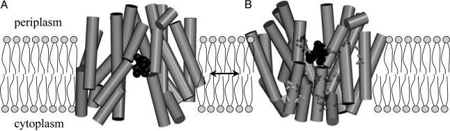
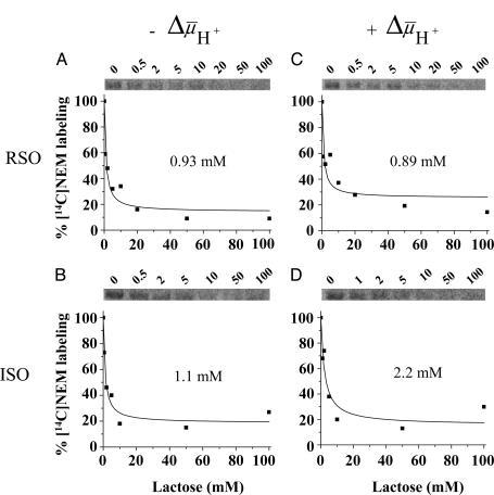
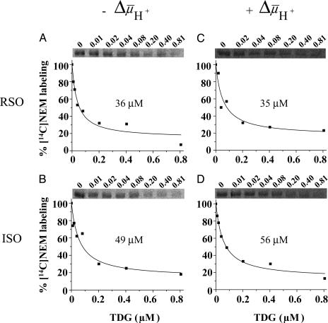
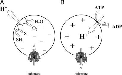
The x-ray structure of lactose permease of Escherichia coli (LacY) exhibits a single sugar-binding site at the apex of a hydrophilic cavity open to the cytoplasm, and it has been postulated that the binding site has alternating access to either side of the membrane during turnover. Here, the affinity of LacY for ligand in right-side-out or inside-out membrane vesicles is measured in the absence or presence of an H(+) electrochemical gradient (Deltamicro(H(+))) by utilizing ligand protection against alkylation. Right-side-out or inside-out membrane vesicles containing LacY with a single cysteine residue at position 148 exhibit K(D) values for lactose or beta-d-galactopyranosyl 1-thio-beta-d-galactopyranoside of approximately 1.0 mM or 40 microM, respectively, and no systematic change is observed in the presence of Deltamicro(H(+)) under conditions in which there is little or no accumulation of ligand. The results are consistent with a mechanism in which the major effect of Deltamicro(H(+)) on sugar accumulation is caused by an increased rate of deprotonation on the inner face of the membrane, leading to an increase in the rate of return of the unloaded symporter to the outer face of the membrane.


Figures

Fig. 1.
Fig. 1.
Postulated structural changes between inward- and outward-facing LacY conformations with bound TDG. (A) Inward-facing conformation viewed parallel to the membrane. (B) A possible model for the outward-facing conformation based on chemical modification and cross-linking experiments, viewed parallel to the membrane. The model was obtained as described in ref. .
Fig. 2.
Fig. 2.
Rate of NEM labeling. Ice-cold RSO vesicles containing single-Cys-148 LacY at 10 mg of protein per ml were added to a solution containing [14C]NEM (40 mCi/mmol, 0.5 mM final concentration). Reactions were carried out on ice and quenched with 10 mM dithiothreitol at the given time. Biotinylated LacY was solubilized in dodecyl β-d-maltopyranoside and purified by affinity chromatography on monomeric avidin Sepharose. Samples were subjected to SDS/12% polyacrylamide gel electrophoresis, and 14C-labeled protein was quantitated with a Typhoon 9410 PhosphorImager (Molecular Dynamics). A.U., arbitrary units.
Fig. 3.
Fig. 3.
Lactose protection against [14C]NEM labeling of single-Cys-148 LacY. (A and B) Ice-cold RSO or ISO vesicles containing single-Cys-148 mutant at 10 mg of protein per ml were added to a solution containing [14C]NEM (40 mCi/mmol, 0.5 mM final concentration) and given concentrations of lactose and incubated on ice for 5 min. Reactions were quenched with 10 mM dithiothreitol, and biotinylated LacY was purified and analyzed as described for Fig. 2. Labeling in the presence of a given concentration of lactose is expressed as percent labeling observed in the absence of ligand. (C and D) Effect of formula image on the lactose protection against Cys-148 alkylation by NEM. Ice-cold RSO or ISO membrane vesicles were added with 20 mM lithium d-lactate under oxygen or 10 mM Mg(II)ATP, respectively, and incubated on ice for 5 min before addition to a solution containing [14C]NEM and given concentrations of lactose as described above (final protein concentration, 10 mg/ml). KD values were calculated as described in Materials and Methods.
Fig. 4.
Fig. 4.
TDG protection against [14C]NEM labeling of single-Cys-148 LacY in RSO or ISO vesicles in the absence and presence of formula image. Experiments were carried out as described for Fig. 3 with given concentrations of TDG. KD values were calculated as described in Materials and Methods.
Fig. 5.
Fig. 5.
Effect of formula image of opposite polarities on substrate translocation in RSO or ISO vesicles. (A) formula image with RSO vesicles generated by addition of d-lactate (ΔΨ, interior negative); substrate is accumulated. (B) formula image with ISO vesicles generated by addition of 10 mM Mg(II)ATP or 20 mM lithium d-lactate under oxygen (interior positive and/or acid), which is opposite to that of RSO vesicles; substrate effluxes from the vesicles.

References

    1. Saier, M. H., Jr. (2000) Mol. Microbiol. 35, 699-710. - PubMed
    1. Kaback, H. R., Sahin-Tóth, M. & Weinglass, A. B. (2001) Nat. Rev. Mol. Cell Biol. 2, 610-620. - PubMed
    1. Schuldiner, S. & Kaback, H. R. (1977) Biochim. Biophys. Acta 472, 399-418. - PubMed
    1. Frillingos, S. & Kaback, H. R. (1996) Biochemistry 35, 3950-3956. - PubMed
    1. Lolkema, J. S., Carrasco, N. & Kaback, H. R. (1991) Biochemistry 30, 1284-1290. - PubMed

Publication types

MeSH terms

LinkOut - more resources