. 2004 Aug;53(4):1099-108.
doi: 10.1111/j.1365-2958.2004.04192.x.

Silencing the ap65 gene reduces adherence to vaginal epithelial cells by Trichomonas vaginalis

Affiliations

Silencing the ap65 gene reduces adherence to vaginal epithelial cells by Trichomonas vaginalis

V Mundodi et al. Mol Microbiol. 2004 Aug.

Abstract

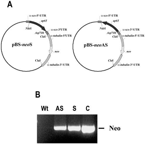
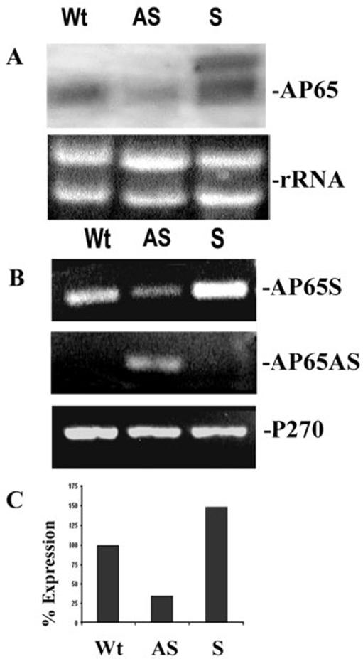
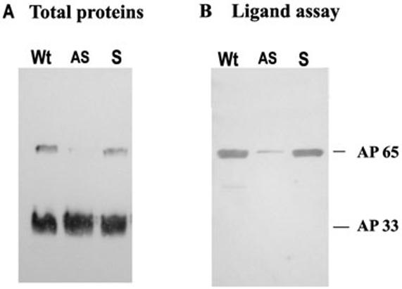
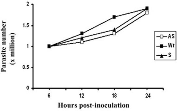
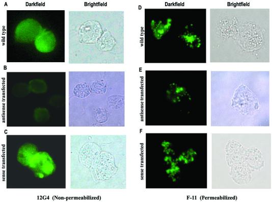
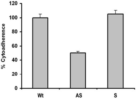
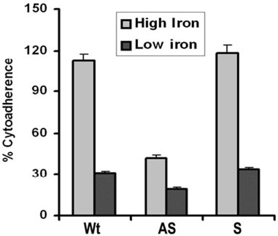
Host parasitism by Trichomonas vaginalis is complex and in part mediated by adherence to vaginal epithelial cells (VECs). Four trichomonad surface proteins bind VECs as adhesins, and AP65 is a major adhesin with sequence identity to an enzyme of the hydrogenosome organelle that is involved in energy generation. In order to perform genetic analysis and assess the role of AP65 in T. vaginalis adherence, we silenced expression of ap65 using antisense RNA. The gene for ap65 was inserted into the vector pBS-neo in sense and antisense orientations to generate plasmids pBS-neoS (S) and pBS-neoAS (AS), respectively. Trichomonads were then transfected with S and AS plasmids for selection of stable transfectants using Geneticin, and the presence of plasmid in transfectants was confirmed by polymerase chain reaction of the neo gene. Reverse transcription polymerase chain reaction and Northern blot analysis showed decreased amounts of ap65 transcript in AS transfected parasites. Growth kinetics of the antisense-transfected and wild type organisms were similar, suggesting that silencing AP65 did not affect overall energy generation for growth. Immunoblot analysis using monoclonal antibody (mAb) to AP65 of AS transfectants showed decreased amounts of AP65 when compared to wild type or S transfectants. Not unexpectedly, this corresponded to decreased amounts of AP65 bound to VECs in a functional ligand assay. Reduction in parasite surface expression of AP65 was related to lower levels of adherence to VECs by AS-transfectants compared to control organisms. Antisense silencing of ap65 was not alleviated by growth of trichomonads in high iron, which up-regulates transcription of ap65. Our work reaffirms the role for AP65 as an adhesin, and in addition, we demonstrate antisense RNA gene silencing in T. vaginalis to study the contribution of specific genes in pathogenesis.


Figures

Fig. 1
Fig. 1
Constructs for expression of ap65 sense (S) and antisense (AS) RNA (A) and PCR amplification of the neo coding region in transfected parasites (B) to demonstrate the presence of plasmids. Part A shows the individual plasmids with the ap65-3 gene in the sense (left plasmid) vs. the antisense (right) orientation. The parent plasmid pBS-FDHAHA-2neo with the 5′- and 3′-α-succinyl coA synthetase untranslated region (UTR) and the neomycin (neo) gene flanked by the 5′- and 3′-α-tubulin UTR was engineered to carry the ap65-3 open reading frame in the S and AS orientations using the NdeI and ASP718 restriction sites. A description of the origin of the plasmid is described in Experimental procedures. Part B presents results of a PCR reaction to amplify the 795-bp neo gene from DNA derived from trichomonads transfected by electroporation with the respective plasmids (lanes labelled AS and S) and compared to the PCR product from the plasmid alone as control (lane C). Total genomic DNA was used as template for the PCR reactions using S and AS primers specific to the 795-bp neo coding region. The lane labelled Wt is of control wild type parasites to confirm the absence of plasmid or cross-hybridizing DNA.
Fig. 2
Fig. 2
Levels of ap65 mRNA in antisense (AS) transfected trichomonads is less than transcript in the sense (S) and wild type (Wt) organisms. A. Northern analysis was performed to detect levels of ap65 mRNA. The larger band in the S transfectants is of the ap65 transcript derived from the S plasmid. Using the Scion image β program and densitometric scanning of the bands indicates a 62% reduction of transcript in the AS transfectants. The rRNA bands are included as controls to show equivalent amounts of RNA added to the blots; however, the slightly increased intensity of the S ap65 transcript is the result of slightly higher amounts of sample added to the well. In this experiment, 10 μg total RNA in each lane was electrophoresed in 1.2% agarose-formaldehyde gels and blotted onto Hybond-P membranes. The blot was probed with DIG-labelled ap65 that hybridized to an approximately 2-kb endogenous ap65 transcript. B. A representative experiment showing RT-PCR products for the ap65 sense transcript (AP65S) and the episomally expressed antisense transcript (AP65AS). RT-PCR was also performed using primers to amplify a 300-bp region of the p270 gene (P270) as a control. Equal volumes of the PCR reactions were separated on 1% agarose followed by staining with ethidium bromide. C. Quantitation of amounts of PCR products in part A for the ap65 transcript in AS transfected parasites compared to S transfected and Wt organisms. The bar graph shows the relative amounts of the RT-PCR products for ap65. The amount of Wt ap65 transcript was normalized to 100%. As for Part A, quantitation was done using the Scion image β program and densitometric scanning.
Fig. 3
Fig. 3
Immunoblots with specific mAb DM116 detecting the amount of AP65 in total protein preparations (A) and bound to HeLa cells in a ligand assay (B) from equal numbers of S and AS transfected trichomonads compared to control Wt organisms. A. Total proteins from 107 trichomonads were subjected to SDS-PAGE on 10% acrylamide before blotting onto Hybond-P membranes. As a control to show equivalent protein amounts in each lane, the blot was also probed with mAb F5 to the AP33 adhesin (Engbring and Alderete, 1998a; 1998b). B. AP65 bound to HeLa cells were solubilized and electrophoresed for blotting and probing with mAb DM116 as in part A. Total proteins and the ligand assay were as described in Experimental procedures.
Fig. 4
Fig. 4
Growth kinetics of wild type and transfected trichomonads. The starting density was 105 organisms. Numbers of parasites were enumerated using a Neubauer hemocytometer at the different time points. The results from four different growth experiments were averaged, and cell numbers did not differ by more than 5% of the values given.
Fig. 5
Fig. 5
Immunofluorescence and corresponding brightfield microscopy showing decreased surface (non-permeabilized) and non-surface (permeabilized) AP65 in antisense transfected (B and E) compared to wild type (A and D) and sense transfected (C and F) trichomonads. The fluorescence patterns seen in permeabilized organisms are expected and represent the protein within the hydrogenosome organelles.
Fig. 6
Fig. 6
Antisense (AS) transfected T. vaginalis organisms have lower levels of adherence to immortalized MS-74 vaginal epithelial cells compared to sense transfected (S) and wild type (Wt) trichomonads. The extent of adherence by Wt parasites was normalized to 100% for comparative purposes, as before (Arroyo et al., 1992; Garcia et al., 2003). The results are the average and standard deviations from four different experiments. Each experiment was performed with quadruplicate samples.
Fig. 7
Fig. 7
High iron does not restore levels of adherence in antisense transfected T. vaginalis organisms to those seen for Wt and S transfected trichomonads. The extent of adherence by Wt parasites grown in iron-replete medium was normalized to 100% for comparative purposes. The results are the average and standard deviations from four different experiments, and each experiment was performed with quadruplicate samples.

References

    1. Alderete JF. Identification of immunogenic and antibody-binding proteins on the membrane of pathogenic Trichomonas vaginalis. Infect Immun. 1983;40:284–291. - PMC - PubMed
    1. Alderete JF, Demeš P, Gombošova A, Valent M, Fabušova M, Janoœka A, et al. Specific parasitism of purified vaginal epithelial cells by Trichomonas vaginalis. Infect Immun. 1988;56:2558–2562. - PMC - PubMed
    1. Alderete JF, Engbring J, Lauriano CM, O’Brien JL. Only two of the Trichomonas vaginalis triplet AP51 adhesins are regulated by iron. Microb Pathog. 1998;23:1–16. - PubMed
    1. Alderete JF, Garza GE. Specific nature of Trichomonas vaginalis parasitism of host cell surfaces. Infect Immun. 1985;50:701–708. - PMC - PubMed
    1. Alderete JF, Millsap KW, Lehker MW, Benchimol M. Enzymes on microbial pathogens and Trichomonas vaginalis: molecular mimicry and functional diversity. Cell Microbiol. 2001;3:1–13. - PubMed

Publication types

Associated data