

Nuclear cloning of embryonal carcinoma cells
- PMID: 15306687
- PMCID: PMC521109
- DOI: 10.1073/pnas.0405015101
Nuclear cloning of embryonal carcinoma cells
Erratum in
- Proc Natl Acad Sci U S A. 2004 Sep 28;101(39):14305
Abstract
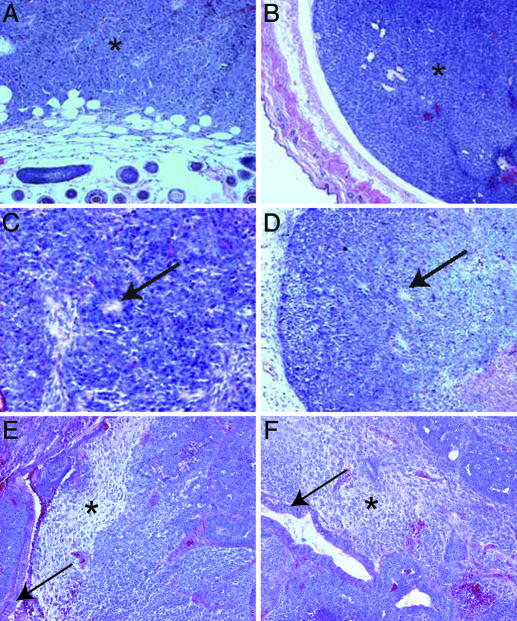
Embryonal carcinoma (EC) cells have served as a model to study the relationship between cancer and cellular differentiation given their potential to produce tumors and, to varying degrees, participate in embryonic development. Here, nuclear transplantation was used to assess the extent to which the tumorigenic and developmental potential of EC cells is governed by epigenetic as opposed to genetic alterations. Nuclei from three independent mouse EC cell lines (F9, P19, and METT-1) with differing developmental and tumorigenic potentials all were able to direct early embryo development, producing morphologically normal blastocysts that gave rise to nuclear transfer (NT)-derived embryonic stem (ES) cell lines at a high efficiency. However, when tested for tumor or chimera formation, the resulting NT ES cells displayed an identical potential as their respective donor EC cells, in stark contrast to previously reported NT ES cells derived from transfer of untransformed cells. Consistent with this finding, comparative genomic hybridization identified previously undescribed genetic lesions in the EC cell lines. Therefore, nonreprogrammable genetic modifications within EC nuclei define the developmental and tumorigenic potential of resulting NT ES cells. Our findings support the notion that cancer results from the deregulation of stem cells and further suggest that the genetics of ECs will reveal genes involved in stem cell self-renewal and pluripotency.
Figures


References
-
- Jaenisch, R. & Bird, A. (2003) Nat. Genet. 33, Suppl., 245–254. - PubMed
-
- Li, E. (2002) Nat. Rev. Genet. 3, 662–673. - PubMed
-
- Jones, P. A. & Baylin, S. B. (2002) Nat. Rev. Genet. 3, 415–428. - PubMed
-
- Herman, J. G. & Baylin, S. B. (2003) N. Engl. J. Med. 349, 2042–2054. - PubMed
-
- Hochedlinger, K. & Jaenisch, R. (2002) Curr. Opin. Cell Biol. 14, 741–748. - PubMed
Publication types
MeSH terms
Grants and funding
LinkOut - more resources
Full Text Sources
Other Literature Sources
Research Materials
Miscellaneous
