Comparative Study
. 2004 Sep 1;23(17):3462-71.
doi: 10.1038/sj.emboj.7600357. Epub 2004 Aug 12.

Structural basis for proteolysis-dependent activation of the poliovirus RNA-dependent RNA polymerase

Affiliations
Comparative Study

Structural basis for proteolysis-dependent activation of the poliovirus RNA-dependent RNA polymerase

Aaron A Thompson et al. EMBO J. .

Abstract

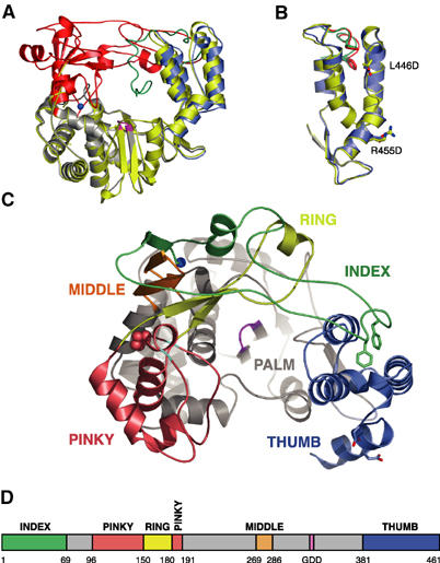
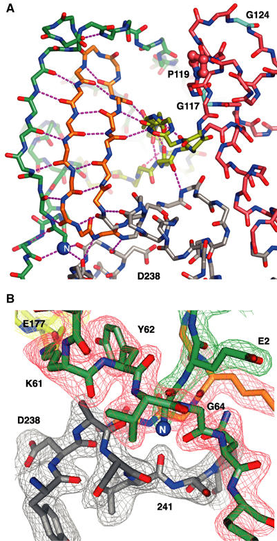
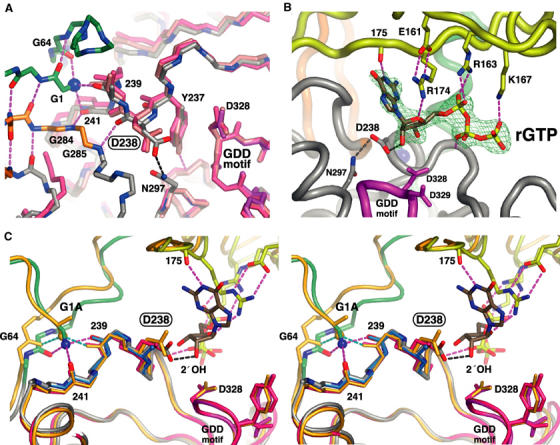
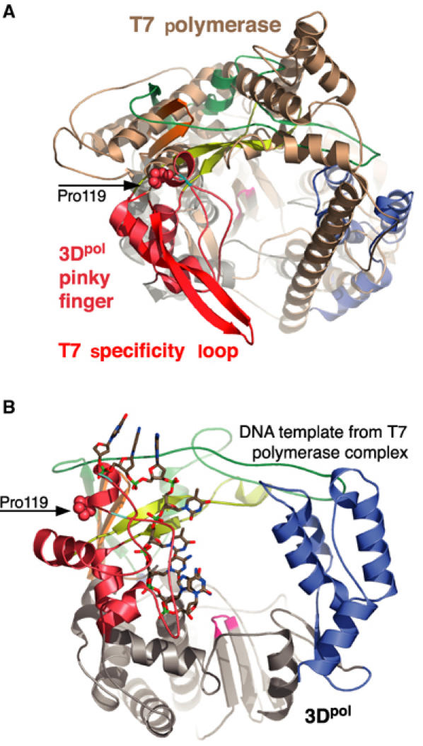
The active RNA-dependent RNA polymerase of poliovirus, 3Dpol, is generated by cleavage of the 3CDpro precursor protein, a protease that has no polymerase activity despite containing the entire polymerase domain. By intentionally disrupting a known and persistent crystal packing interaction, we have crystallized the poliovirus polymerase in a new space group and solved the complete structure of the protein at 2.0 A resolution. It shows that the N-terminus of fully processed 3Dpol is buried in a surface pocket where it makes hydrogen bonds that act to position Asp238 in the active site. Asp238 is an essential residue that selects for the 2' OH group of substrate rNTPs, as shown by a 2.35 A structure of a 3Dpol-GTP complex. Mutational, biochemical, and structural data further demonstrate that 3Dpol activity is exquisitely sensitive to mutations at the N-terminus. This sensitivity is the result of allosteric effects where the structure around the buried N-terminus directly affects the positioning of Asp238 in the active site.


Figures

Figure 1
Figure 1
Overview of poliovirus 3Dpol RdRp structure. (A) Comparison of the original partial structure (yellow) with the complete structure shown with the fingers domain in red, the palm in gray, the thumb in blue, and the active site colored magenta. The N-terminal strand (residues 12–36) of the original structure that descended toward the active site is shown in green. The two structures were superimposed using the backbone atoms of the active site GDD motif and three residues on either side of it (i.e. residues 324–332). (B) Superimposition of the thumb domains from the original structure (yellow) and new complete structure (blue) showing that the thumb structure is largely unchanged by the two mutations (L446D and R455D) used to break Interface I and crystallize 3Dpol in a new lattice. The side chains of Phe30 and Phe34 are shown in green for the original structure and red for the new complete structure. (C) Top view of the complete 3Dpol structure highlighting the individual fingers of the fingers domain. The index finger is shown in green, the middle finger in orange, the ring finger in yellow, and the pinky finger in pink. As in (A), the palm is shown in gray, the thumb is in blue, and the active site is colored magenta. Phe30 and Phe34 are shown as sticks, Pro119 on the pinky finger is indicated with spheres, and glycines 117 and 124 are colored in cyan. (D) Bar representation of the 3Dpol sequence colored according to the structural elements shown in (C). Sections of the sequence in the palm are in gray and the numbers correspond to the first residue in a given structural motif.
Figure 2
Figure 2
Structural details of the 3Dpol fingers domain and buried N-terminus. (A) Structure of the fingers domain highlighting the extensive network of hydrogen bonds linking the N-terminus (blue sphere at lower left) and N-terminal strand of the index finger to the middle and ring fingers. Note the putative template entry channel separating pinky finger (pink carbon atoms) from the rest of the fingers domain and how the ring finger (yellow) is an insertion in the pinky finger structure. Proline 119 and glycines 117 and 124 that may play a role in template binding are highlighted (see Discussion). The view is from the left side of Figure 1A and C. (B) Electron density map of the region surrounding the buried N-terminus. The map is a 2.0 Å resolution simulated annealing (1500 K) composite omit 2FoFc map contoured at 1.6σ. The view is from the left side as compared to (A), the carbon atoms of the various structural motifs are colored as in Figure 1C and D, and corresponding sections of the density map are colored differently for clarity.
Figure 3
Figure 3
Molecular details of the 3Dpol nucleotide-binding site illustrating how the buried N-terminus positions Asp238 for interactions with the 2′ OH group of the bound NTP. (A) Superposition of three 3Dpol structures showing the selective ∼1.4 Å movement of Asp238 toward the active site when the N-terminus is properly positioned. The original partial wild-type structure is in pink, the 3Dpol Δ68/L446A/R455D structure is in salmon, and the complete structure is colored by atom type with carbons colored according to structural motifs as in Figure 1C. Most side chains have been omitted for clarity and residues 324–332 of the active site (magenta) were used for the superimpositions. (B) Electron density map and model of the GTP molecule bound to 3Dpol with the 2′ OH group making a 2.8 Å long hydrogen bond with Asp238. The GTP makes bridging interactions between the fingers and palm domains. The base is staked on Arg174 from the ring finger, the ribose interacts with Arg174 from the ring finger and Asp238 in the palm, and the triphosphate interacts with Arg163 and Lys167 from the ring finger and the backbone of the palm domain. The map is a 2.35 Å resolution 2FoFc simulated annealing (1500 K) composite omit map contoured at 1.6σ around the rGTP molecule bound after soaking crystals in 10 mM GTP. (C) Stereo view showing how the buried N-terminus of 3Dpol positions Asp238 for rNTP interactions. The N-terminus forms three hydrogen bonds with the carbonyl oxygens of residues 64, 239, and 241 (magenta bonds) that act to position Asp238 for interaction with the 2′ OH of rNTPs. The structures of the G1A mutant (orange), D238A mutant (teal, only residues 238–241 are shown), and original partial structure without a buried N-terminus (red) are superimposed using the active site.
Figure 4
Figure 4
Comparison of the poliovirus 3Dpol and bacteriophage T7 polymerases based on superimposing the Cα atoms of their ‘motif C' structures. This motif contains the pair of β-strands in the core of the palm that present the β-turn GDD motif in the active site. (A) Superposition with the T7 protein structure (brown) from the initiation complex (1CEZ) showing the structural alignment of the 3Dpol pinky finger with the T7 specificity loop (red). (B) Position of the DNA template strand from the T7 elongation complex (1MSW) after the motif C superimposition showing how this predicted template strand path in 3Dpol would collide with the helix/loop containing the cis Pro119 residue (spheres) flanked by glycines 117 and 124 (cyan).

References

    1. Ago H, Adachi T, Yoshida A, Yamamoto M, Habuka N, Yatsunami K, Miyano M (1999) Crystal structure of the RNA-dependent RNA polymerase of hepatitis C virus. Struct Fold Des 7: 1417–1426 - PubMed
    1. Andino R, Rieckhof GE, Achacoso PL, Baltimore D (1993) Poliovirus RNA synthesis utilizes an RNP complex formed around the 5′-end of viral RNA. EMBO J 12: 3587–3598 - PMC - PubMed
    1. Andreotti AH (2003) Native state proline isomerization: an intrinsic molecular switch. Biochemistry 42: 9515–9524 - PubMed
    1. Arnold JJ, Cameron CE (2000) Poliovirus RNA-dependent RNA polymerase 3D(pol). Assembly of stable, elongation-competent complexes by using a symmetrical primer–template substrate (sym/sub). J Biol Chem 275: 5329–5336 - PubMed
    1. Brandts JF, Halvorson HR, Brennan M (1975) Consideration of the possibility that the slow step in protein denaturation reactions is due to cis–trans isomerism of proline residues. Biochemistry 14: 4953–4963 - PubMed

Publication types

MeSH terms