. 2004 Sep 1;23(17):3472-82.
doi: 10.1038/sj.emboj.7600358. Epub 2004 Aug 12.

Mss51p and Cox14p jointly regulate mitochondrial Cox1p expression in Saccharomyces cerevisiae

Affiliations

Mss51p and Cox14p jointly regulate mitochondrial Cox1p expression in Saccharomyces cerevisiae

Antoni Barrientos et al. EMBO J. .

Abstract

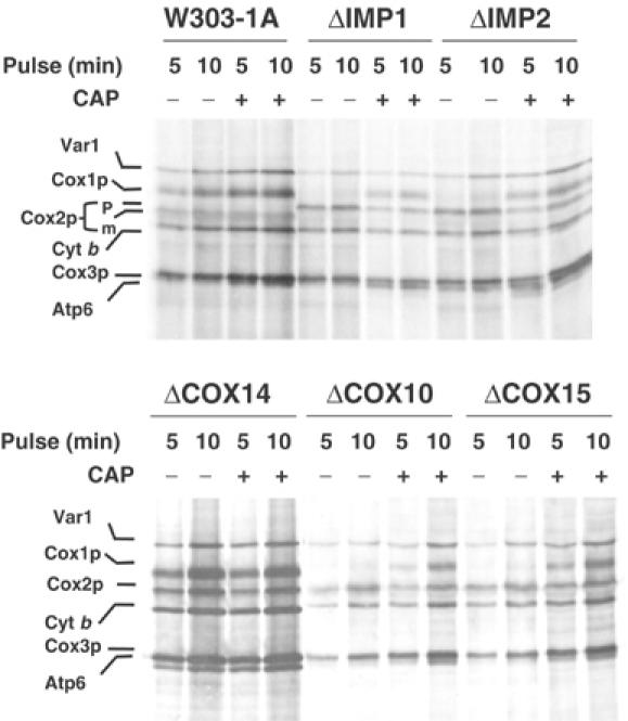
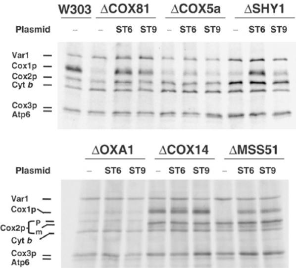
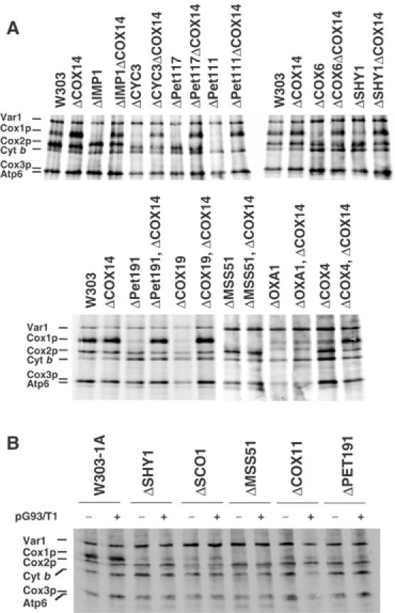
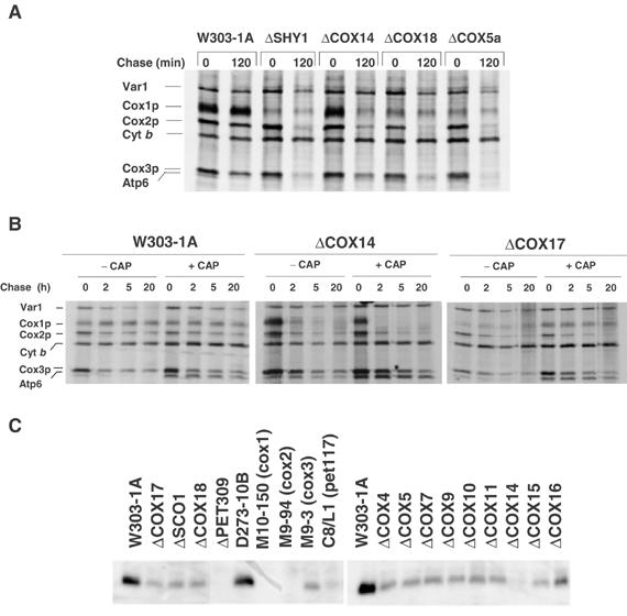
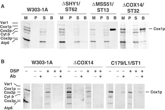
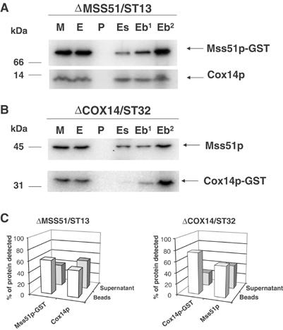
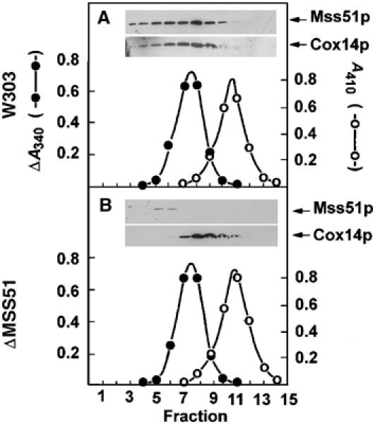
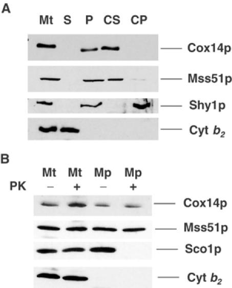
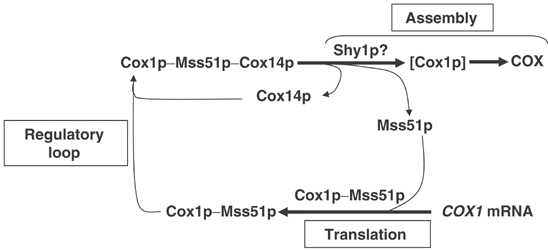
Mutations in SURF1, the human homologue of yeast SHY1, are responsible for Leigh's syndrome, a neuropathy associated with cytochrome oxidase (COX) deficiency. Previous studies of the yeast model of this disease showed that mutant forms of Mss51p, a translational activator of COX1 mRNA, partially rescue the COX deficiency of shy1 mutants by restoring normal synthesis of the mitochondrially encoded Cox1p subunit of COX. Here we present evidence showing that Cox1p synthesis is reduced in most COX mutants but is restored to that of wild type by the same mss51 mutation that suppresses shy1 mutants. An important exception is a null mutation in COX14, which by itself or in combination with other COX mutations does not affect Cox1p synthesis. Cox14p and Mss51p are shown to interact with newly synthesized Cox1p and with each other. We propose that the interaction of Mss51p and Cox14p with Cox1p to form a transient Cox14p-Cox1p-Mss51p complex functions to downregulate Cox1p synthesis. The release of Mss51p from the complex occurs at a downstream step in the assembly pathway, probably catalyzed by Shy1p.


Figures

Figure 1
Figure 1
In vivo labeling of mitochondrial gene products in COX mutants. Wild-type (W303-1A) and mutant cells (described in Table I) were labeled with [35S]methionine at 30°C for the indicated times in the presence of cycloheximide. One-half of each culture was incubated in the presence of 2 mg/ml chloramphenicol during the last 2 h of growth prior to labeling (+CAP). Samples were removed after the indicated times of labeling and processed as detailed in Materials and methods. The mitochondrial translation products are identified in the margin. Cox2p is not processed in ΔIMP1 and ΔIMP2 mutants. The Cox2p precursor (p) in these strains migrates slower that the mature Cox2p (m).
Figure 2
Figure 2
In vivo labeling of mitochondrial gene products in COX mutants expressing different alleles of MSS51. MSS51 and the suppressor mss51T167R, which partially suppress the respiratory defect of shy1 mutants, were cloned in YIp351. The resultant constructs pSG91/ST9 and pSG91/ST6, respectively, were integrated at the chromosomal LEU2 locus of the indicated mutants (see Table I for description of mutants). The mutants (−) and transformants were labeled with [35S]methionine at 30°C for 15 min in the presence of cycloheximide.
Figure 3
Figure 3
In vivo labeling of mitochondrial gene products in COX14 mutants and effect of overexpression of COX14p on Cox1p labeling. (A) Wild type (W303), different COX mutants, and the same mutants carrying an additional null mutation in COX14 were labeled with [35S]methionine at 30°C for 15 min in the presence of cycloheximide. With the exception of CYC3, which codes for a cytochrome c-specific heme lyase, the functions affected in the different strains are described in Table I. (B) The wild-type W303-1A, the indicated COX mutants, and the same strains harboring COX14 on a multicopy plasmid (pG93/T1) were pulse-labeled with [35S]methionine and equivalent amounts of protein were separated by SDS–PAGE on a 17.5% polyacrylamide gel.
Figure 4
Figure 4
Turnover of in vivo-labeled mitochondrial translation products and steady-state concentration of Cox1p in wild type and COX mutants. (A) Wild type (W303-1A) and mutants (described in Table I) were grown and labeled for 20 min at 30°C with [35S]methionine. Labeling was terminated by addition of 80 μmol cold methionine and 12 μg/ml puromycin (0 time). Samples of the cultures were collected after the indicated times of incubation at 30°C and processed as in Figure 1. (B) The wild-type strain W303-1A and the cox14 and cox17 mutants were labeled and chased for the indicated times as in panel A. One-half of each culture was incubated in the presence of chloramphenicol as in Figure 1 prior to labeling. (C) The wild-type W303-1A and D273-10B and mutant strains were grown in 2% galactose, 1% yeast extract, and 2% peptone to stationary phase. Mitochondria were prepared and 10 μg of protein was separated by SDS–PAGE on a 12% polyacrylamide gel. The proteins were transferred to nitrocellulose and probed with a polyclonal antibody against yeast Cox1p. The antibody–antigen complexes were visualized by a secondary reaction with [125I]protein A.
Figure 5
Figure 5
The cox14 defect is not rescued by a ρ genome with COX1 fused to the 5′-UTR of COB. (A) Map of the mitochondrial bypass suppressor ρSUP2. The rearrangement in the suppressor leads to a fusion of the 5′-UTR of COB to nucleotide −174 of COX1 (Manthey and McEwen, 1995). (B) ρSUP2 was transferred by cytoduction to a kar1 mutant (Conde and Fink, 1976) lacking mitochondrial DNA (ρ°). The SUP2 suppressor was transferred from the kar1 donor to ρ° derivatives of null mutants of PET309, MSS51, COX14, and SHY1. The different mutants with the SUP2 genome (ρSUP2) were then crossed to the isogenic mutants with wild-type mitochondrial DNA (ρ+) to obtain the heteroplasmic diploid mutants of PET309 (a/α-ΔPET309/ρ+ρSUP2), MSS51 (a/α-ΔMSS51/ρ+ρSUP2), COX14 (a/α-ΔCOX14309/ρ+ρSUP2), and SHY1 (a/α-ΔSHY1/ρ+ρSUP2). Serial dilutions of the haploid mutants with wild-type mitochondrial DNA and of the diploid strains with the wild-type and suppressor genomes were spotted on YPD and YPEG plates and incubated at 30°C for 2.5 days.
Figure 6
Figure 6
Cox14p and Mss51p interact with Cox1p. (A) Mitochondria were prepared from the wild-type W303-1A, a shy1 null mutant (ΔSHY1/ST62) with a chromosomally integrated plasmid expressing the Shy1p-GST fusion protein, an mss51 null mutant (ΔMSS51/ST13) with a chromosomally integrated plasmid expressing Mss51p-GST, and a cox14 null mutant (ΔCOX14/ST32) with a chromosomally integrated plasmid expressing Cox14p-GST. Mitochondria were labeled with [35S]methionine for 30 min and extracted with 1% lauryl maltoside, 1 M KCl, and 1 mM PMSF. The extract was clarified by centrifugation at 50 000 gav for 30 min and incubated with glutathione–Sepharose beads for 4 h at 4°C. After centrifugation at 1500 rpm for 5 min, the supernatant was collected and the beads were washed three times with PBS. Mitochondria (M) corresponding to 2 μg protein, equivalent volumes of the membrane pellet (P) after lauryl maltoside extraction and of the supernatant from the glutathione–Sepharose beads (S) were separated on a 17.5% polyacrylamide gel by SDS–PAGE. The amount of washed beads (B), however, corresponded to ∼500 μg of the starting mitochondria. (B) Mitochondria from W303-1A, the cox14 null mutant (ΔCOX14), and a cox14 point mutant transformed with a high-copy plasmid containing COX14 (C179/L1/ST1) were labeled for 30 min at 30°C in the presence of [35S]methionine. After a 5 min pulse, the samples were treated with the crosslinker DSP (+) or were mock-treated (−) as described (Hell et al, 2000). Immunoprecipitation of crosslinked adducts was performed using antiserum specific for Cox14p (+) and preimmune serum (−). Immunoprecipitates were analyzed by SDS–PAGE and autoradiography as in Figure 1.
Figure 7
Figure 7
Cox14p interacts with Mss51p. (A) Mitochondria (M) from an mss51 null mutant with a chromosomally integrated plasmid expressing Mss51p-GST fusion protein (ΔMSS51/ST13) were extracted with 1% lauryl maltoside, 1 M KCl, and 1 mM PMSF. The pellet (P) after centrifugation at 50 000 gav for 30 min was suspended in the starting volume of buffer and the extract (E) was mixed and incubated for 4 h at 4°C with glutathione–Sepharose. The supernatants (Es) from the beads were collected and the beads (Eb) were washed three times with PBS. The different fractions adjusted for volume were separated by SDS–PAGE. The lane labeled (Eb2) was loaded with two times the amount of beads. Cox14p and Mss51p-GST were detected by Western blot analysis using specific antibodies against each protein. The proteins were visualized by a secondary reaction with [125I]protein A and the radiolabeled bands were detected with a PhosphorImager (Molecular Dynamics). (B) Same as (A) except that the mitochondria were prepared from a cox14 null mutant with an integrated plasmid expressing a Cox14p-GST fusion protein (ΔCOX14/ST32). (C) The bands shown in (A, B) were quantified with the PhosphorImager. The open bars represent the percentage of the corresponding protein bound to the beads, and the filled bars represent the percentage of unbound protein recovered in the supernatant fraction.
Figure 8
Figure 8
Sedimentation of Mss51p and Cox14p in sucrose gradients. (A) Mitochondria of the wild-type strain W303-1A were extracted at a protein concentration of 10 mg/ml with 1% lauryl maltoside, 20 mM Tris–HCl (pH 7.5), and 0.5 M KCl. The extract (0.4 ml) was mixed with 2.5 mg of hemoglobin and 60 μg lactate dehydrogenase and applied to 4.6 ml of a linear 7–25% sucrose gradient containing 10 mM Tris–HCl and 0.1% Triton X-100. Following centrifugation at 65 000 rpm in a Beckman SW65Ti rotor for 6 h, 14.5 fractions were collected, separated on a 12% polyacrylamide gel, transferred to nitrocellulose and probed with rabbit antiserum against Mss51p or Cox14p followed by a secondary goat peroxidase-conjugated antibody against rabbit IgG. Antibody–antigen complexes were visualized with the Super Signal reagent (Pierce Chemical Co., Rockford, IL). Hemoglobin (○- - -○) was estimated from absorbance at 410 nm and lactate dehydrogenase (⧫- -⧫) was assayed by measuring oxidation of NADH at 340 nm with pyruvate as the substrate. (B) Same as (A) except that the mitochondria were isolated from the mss51 null mutant aW303ΔMSS51 and the gradient was collected in 15 fractions.
Figure 9
Figure 9
Localization and topology of Cox14p and Mss51p. (A) Mitochondria and the post-mitochondrial supernatant fractions were prepared from the wild-type strain W303-1A. A sample of mitochondria at 4 mg/ml was sonically irradiated and centrifuged at 50 000 gav for 30 min. The membrane pellet was suspended in the starting volume of buffer. To 500 μl of the membranes at a protein concentration of 1 mg/ml was added 50 μl of 1 M Na2CO3 (pH 11.3) and 50 mM EDTA. After 30 min on ice, the sample was centrifuged at 100 000 gav for 15 min at 4°C to separate the soluble from the insoluble intrinsic membrane proteins. Equivalent volumes of mitochondria (Mt), membranes (P), the supernatant obtained after centrifugation of the sonicated mitochondria (S), the carbonate supernatant (CS), and pellet (CP) were separated on a 12% polyacrylamide gel, transferred to nitrocellulose, and treated with antiserum against Cox14p as in Figure 8. Antibodies against Mss51p, Shy1p, and cytochrome b2 (Cyt b2) were used to monitor the conversion of mitochondria to mitoplasts and the intactness of the latter. (B) Mitochondria from W303-1A (Glick and Pon, 1995) at a protein concentration of 8 mg/ml in 0.6 M sorbitol and 20 mM Hepes (pH 7.5) (SH) were converted to mitoplasts (Mp) by dilution with eight volumes of 20 mM Hepes (pH 7.5). For controls, mitochondria (Mt) were diluted with eight volumes of SH. Proteinase K (prot K) was added to one-half of each sample at a final concentration of 100 μg/ml. After 60 min on ice, the reaction was stopped by addition of PMSF to a final concentration of 2 mM and the mitochondria and mitoplasts were recovered by centrifugation at 100 000 gav. The pellets were suspended in SH, and proteins were precipitated by addition of 0.1 volume of 50% trichloroacetic acid and heated for 10 min at 65°C. Mitochondrial and mitoplast proteins were separated by SDS–PAGE on a 12% polyacrylamide gel, transferred to nitrocellulose, and probed with antibody against Cox14p, Sco1p, Mss51p, and cytochrome b2. Antibody–antigen complexes were visualized as in (A).
Figure 10
Figure 10
Model depicting Cox1p expression coupled to Shy1p-dependent cytochrome oxidase assembly. Mss51p promotes initiation and elongation of Cox1p translation (see Discussion). Cox14p and Mss51p form a ternary complex with newly synthesized Cox1p. The release of Mss51p from the ternary complex depends on a downstream Cox1p assembly step(s) perhaps involving Shy1p. According to this scheme, mutations blocking assembly trap Mss51p in the ternary complex, thereby limiting its availability for translation. In the cox14 mutant, Mss51p is still able to complex with Cox1p. The resultant binary complex is not assembly-competent causing Cox1p to be diverted to degradation with a concomitant release of Mss51p for additional rounds of Cox1p synthesis.

References

    1. Aldridge P, Hughes KT (2002) Regulation of flagellar assembly. Curr Opin Microbiol 5: 160–165 - PubMed
    1. Barrientos A, Barros MH, Valnot I, Rotig A, Rustin P, Tzagoloff A (2002a) Cytochrome oxidase in health and disease. Gene 286: 53–63 - PubMed
    1. Barrientos A, Korr D, Tzagoloff A (2002b) Shy1p is necessary for full expression of mitochondrial COX1 in the yeast model of Leigh's syndrome. EMBO J 21: 43–52 - PMC - PubMed
    1. Barros MH, Carlson CG, Glerum DM, Tzagoloff A (2001) Involvement of mitochondrial ferredoxin and Cox15p in hydroxylation of heme O. FEBS Lett 492: 133–138 - PubMed
    1. Barros MH, Tzagoloff A (2002) Regulation of the heme A biosynthetic pathway in Saccharomyces cerevisiae. FEBS Lett 516: 119–123 - PubMed

Publication types

MeSH terms