. 2004 Aug 12:5:109.
doi: 10.1186/1471-2105-5-109.

The Hotdog fold: wrapping up a superfamily of thioesterases and dehydratases

Affiliations

The Hotdog fold: wrapping up a superfamily of thioesterases and dehydratases

Shane C Dillon et al. BMC Bioinformatics. .

Abstract

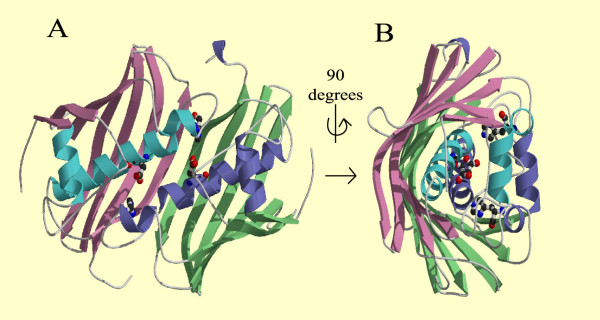
Background: The Hotdog fold was initially identified in the structure of Escherichia coli FabA and subsequently in 4-hydroxybenzoyl-CoA thioesterase from Pseudomonas sp. strain CBS. Since that time structural determinations have shown a number of other apparently unrelated proteins also share the Hotdog fold.

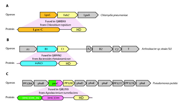
Results: Using sequence analysis we unify a large superfamily of HotDog domains. Membership includes numerous prokaryotic, archaeal and eukaryotic proteins involved in several related, but distinct, catalytic activities, from metabolic roles such as thioester hydrolysis in fatty acid metabolism, to degradation of phenylacetic acid and the environmental pollutant 4-chlorobenzoate. The superfamily also includes FapR, a non-catalytic bacterial homologue that is involved in transcriptional regulation of fatty acid biosynthesis. We have defined 17 subfamilies, with some characterisation. Operon analysis has revealed numerous HotDog domain-containing proteins to be fusion proteins, where two genes, once separate but adjacent open-reading frames, have been fused into one open-reading frame to give a protein with two functional domains. Finally we have generated a Hidden Markov Model library from our analysis, which can be used as a tool for predicting the occurrence of HotDog domains in any protein sequence.

Conclusions: The HotDog domain is both an ancient and ubiquitous motif, with members found in the three branches of life.


Figures

Figure 1
Figure 1
The structure of the active HotDog domain dimer. (A) A ribbon representation of the Escherichia coli FabA dimer (PDB code: 1MKB), viewed along the dyad axis. Each 171-residue subunit contains a Hotdog fold/ domain, consisting of a seven-stranded antiparallel b-sheet 'bun', coloured magenta and green, and a five-turn a-helical 'sausage' coloured blue and purple in the respective subunits. The Hotdog fold is best observed in Figure B. There are two independent active sites located between the dimers, the active site residues of His70 from one subunit and Asp84 from the other subunit, represented as a ball-and-stick model with CPK colouring (carbon, black; hydrogen, white; oxygen, red; nitrogen, blue), constitute the potential reactive protein groups in the active sites [1]. (B) A view of FabA rotated 90° along the dyad axis. The figures were generated with MOLSCRIPT [69] and rendered with RASTER3D [70].
Figure 2
Figure 2
A schematic Figure showing the various domain organizations of proteins with a HotDog domain. For each distinct architecture we show an example protein, the species it has come from and its length is shown in parentheses. The key identifies all the domains in the Figure and also includes the Pfam accession number or numbers describing each domain.
Figure 3
Figure 3
Rosetta fusion proteins in the HotDog domain superfamily. For each fusion event we show an example operon containing the two proteins separate and an example of the fused rosetta protein.

References

    1. Leesong M, Henderson BS, Gillig JR, Schwab JM, Smith JL. Structure of a dehydratase-isomerase from the bacterial pathway for biosynthesis of unsaturated fatty acids: two catalytic activities in one active site. Structure. 1996;4:253–264. doi: 10.1016/S0969-2126(96)00030-5. - DOI - PubMed
    1. Benning MM, Wesenberg G, Liu R, Taylor KL, Dunaway-Mariano D, Holden HM. The three-dimensional structure of 4-hydroxybenzoyl-CoA thioesterase from Pseudomonas sp. Strain CBS-3. J Biol Chem. 1998;273:33572–33579. doi: 10.1074/jbc.273.50.33572. - DOI - PubMed
    1. Thoden JB, Zhuang Z, Dunaway-Mariano D, Holden HM. The structure of 4-hydroxybenzoyl-CoA thioesterase from arthrobacter sp. strain SU. J Biol Chem. 2003;278:43709–43716. doi: 10.1074/jbc.M308198200. - DOI - PubMed
    1. Zhuang Z, Song F, Takami H, Dunaway-Mariano D. The BH1999 protein of Bacillus halodurans C-125 is gentisyl-coenzyme A thioesterase. J Bacteriol. 2004;186:393–399. doi: 10.1128/JB.186.2.393-399.2004. - DOI - PMC - PubMed
    1. Li J, Derewenda U, Dauter Z, Smith S, Derewenda ZS. Crystal structure of the Escherichia coli thioesterase II, a homolog of the human Nef binding enzyme. Nat Struct Biol. 2000;7:555–559. doi: 10.1038/76776. - DOI - PubMed

Publication types

LinkOut - more resources