. 2004 Oct 15;560(Pt 2):429-37.
doi: 10.1113/jphysiol.2004.072108. Epub 2004 Aug 12.

Ageing-related changes of connexins and conduction within the sinoatrial node

Affiliations

Ageing-related changes of connexins and conduction within the sinoatrial node

Sandra A Jones et al. J Physiol. .

Abstract

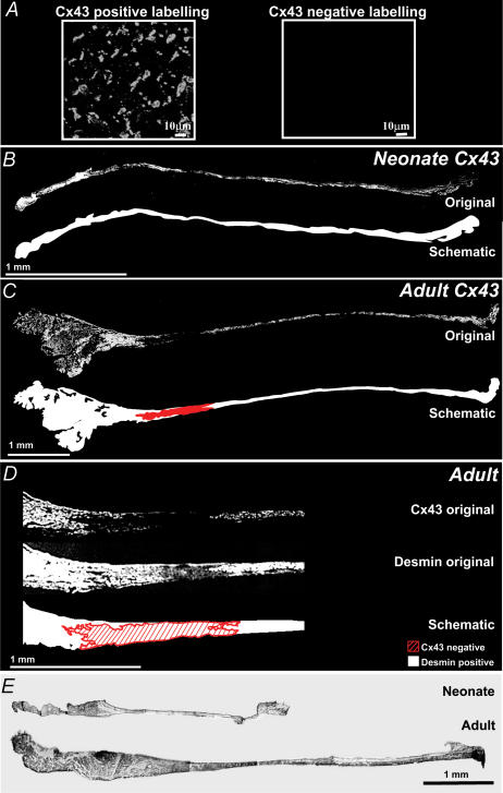
Clinical studies have shown that sinoatrial node dysfunction occurs at the highest incidence in the elderly population. Guinea-pigs were studied throughout their lifespan (i.e. birth to 38 months) to investigate the possible mechanism leading to nodal dysfunction. Using immunofluorescence with confocal microscopy, Cx43 protein expression was shown at birth to be present throughout the sinoatrial node and atrial muscle, however, at one month Cx43 protein was not expressed in the centre of the sinoatrial node. Throughout the remainder of the animal's lifespan the area of tissue lacking Cx43 protein progressively increased. Western blot provided verification by quantitative analysis that Cx43 protein expression within the sinoatrial node decreased with age; however, the expression of other cardiac connexins, Cx40 and Cx45, did not differ with age. Analysis of conduction maps showing propagation of the action potential across the sinoatrial node, from the initiation point to the crista terminalis, found that the action potential conduction time taken and conduction distance increased proportionally with age; conversely the conduction velocity decreased with age. We have shown ageing induces degenerative changes in action potential conduction, contributed to by the observed loss of Cx43 protein. Our data identify Cx43 as a potential therapeutic target for quashing the age-related deterioration of the cardiac pacemaker.


Figures

Figure 1
Figure 1. Physical changes of the guinea-pig during ageing
A, mean body weight (neonate versus young P < 0.0001, young versus adult P < 0.0001), B, mean heart weight (neonate versus young P < 0.0001, young versus adult P < 0.0001) and C, mean heart-to-body weight ratio are shown versus age (n = 5). D, FISH-labelled telomeric DNA from atrial nuclei of different aged animals plus the ‘no probe’ control (scale bar, 1 μm). E, the mean percentage telomere labelling per nuclei versus age (n = 100 nuclei from n = 5 animals; linear regression, y = −3.6049x + 57.04, x is the age in months). Error bars show s.e.m.
Figure 2
Figure 2. Expression of Cx43 protein
A, high magnification examples of positive- and negative-labelled Cx43 protein from atrial tissue are shown. B, an original confocal image of a neonate section labelled with anti-Cx43 antibody showed Cx43 protein was expressed throughout the SA node and surrounding atrial muscle, further illustrated by the schematic image where detected Cx43 protein is shown in white. C, an original confocal image of an adult section labelled with anti-Cx43 antibody identified an area lacking Cx43 protein, shown in the schematic image in red with the remaining tissue expressing Cx43 protein in white. D, high magnification confocal images of sequential serial sections of the SA node region showed the area lacking Cx43 protein was positive for desmin. Desmin protein was continuous and uniformly labelled across the SA node and atrial muscle. A schematic image shows desmin protein in white and the area lacking Cx43-labelled protein hashed in red. E, original images from a light microscope show collagen is present throughout the SA node at all ages studied.
Figure 3
Figure 3. Progressive increase in the area of tissue absent of Cx43 protein during ageing
A, for each age group, schematic images at 0.5 mm intervals are shown on the right and the intact SA node on the left. At each interval, the exact location of the area lacking Cx43 protein has been translated to the intact SA node (SAN) image. The total area where Cx43 protein has not been detected is outlined in red on the intact SA node. B, the mean area of SA node where Cx43 protein expression was not detected is shown versus age. (n = 5; ANOVA P < 0.0001; every group is significantly different from the other P < 0.001). Error bars show s.e.m.
Figure 4
Figure 4. Total content of connexins versus age
A, paired samples of atrial (a) and SA node (s) tissue from animals of different ages were analysed for protein expression. Typical blots for desmin (53 kDa), Cx45 (45 kDa), Cx43 (43 kDa) and Cx40 (40 kDa) are shown. B, Cx43 protein expression in the SA node expressed as a percentage of the mean amount identified in the neonate. Cx43 expression within the SA node significantly fell in a progressive manner correlating with age to reach 2 ± 0.4% of that identified in the neonate animals (n = 5 in each case) by senescence, a 98% decline in Cx43 protein expression within the SA node region. Error bars show s.e.m.
Figure 5
Figure 5. Conduction of the action potential changes with age
A, mean intrinsic heart rate versus age (n = 5; ANOVA P < 0.0001; t test neonate versus young P < 0.0001, young versus adult P < 0.01). BD, typical maps of action potential conduction in SA node preparations from neonate (B), adult (C) and senescent (D) animals. From the position of the leading pacemaker site (*), isochrones are shown at 2 ms intervals to the border of the crista terminalis (solid white line, CT), where the SA node tissue meets the atrial muscle. E–G, mean time taken for conduction of the action potential (E), the distance (F) and velocity from the leading pacemaker site in the SA node to the atrial muscle of the CT (n = 5; time taken, ANOVA P < 0.0001; distance, ANOVA P < 0.0001; velocity, ANOVA P < 0.005).

Similar articles

Cited by

References

    1. Alings AMW, Bouman LN. Electrophysiology of the ageing rabbit and cat sinoatrial node – a comparative study. Eur Heart J. 1993;14:1278–1288. - PubMed
    1. Beardslee MA, Laing JG, Beyer EC, Saffitz JE. Rapid turnover of connexin43 in the adult rat heart. Circ Res. 1998;83:629–635. - PubMed
    1. Di Gennaro M, Bernabei R, Sgadari A, Carosella L, Carbonin PU. Age-related differences in isolated rat sinus node function. Basic Res Cardiol. 1987;82:530–536. - PubMed
    1. Eloff BC, Lerner DL, Yamada KA, Schuessler RB, Saffitz JE, Rosenbaum DS. High resolution optical mapping reveals conduction slowing in connexin43 deficient mice. Cardiovasc Res. 2001;51:681–690. - PubMed
    1. Honjo H, Boyett MR, Coppen SR, Takagishi Y, Opthof T, Severs NJ, Kodama I. Heterogeneous expression of connexins in rabbit sinoatrial node cells: correlation between connexin isotype and cell size. Cardiovasc Res. 2002;53:89–96. - PubMed

Publication types