. 2004 Sep;78(17):9093-104.
doi: 10.1128/JVI.78.17.9093-9104.2004.

Adjuvant activities of novel cytokines, interleukin-23 (IL-23) and IL-27, for induction of hepatitis C virus-specific cytotoxic T lymphocytes in HLA-A*0201 transgenic mice

Affiliations

Adjuvant activities of novel cytokines, interleukin-23 (IL-23) and IL-27, for induction of hepatitis C virus-specific cytotoxic T lymphocytes in HLA-A*0201 transgenic mice

Masanori Matsui et al. J Virol. 2004 Sep.

Abstract

Searching the sequence databases has revealed two novel cytokines: interleukin-23 (IL-23) and IL-27. These cytokines are quite similar to, but clearly distinct from IL-12 in their structures and T-cell stimulatory fashions. In contrast to IL-12, however, little is known about the roles of IL-23 and IL-27 in the immune regulation. Previously, we evaluated the prime-boost immunization consisting of priming and the first boosting with the hepatitis C virus (HCV)-core expression plasmid, followed by a second boosting with recombinant adenovirus expressing HCV core for induction of HCV core-specific cytotoxic T lymphocytes (CTLs) in BALB/c mice. The present study demonstrates that HCV-specific CTL induction was greatly enhanced by coinoculation of an IL-12 expression plasmid in the prime-boost immunization, indicating the potent adjuvant activity of IL-12. We investigated whether similar adjuvant effects could be exerted by either IL-23 or IL-27 in a prime-boost immunization with HLA-A*0201 transgenic mice. Coadministration of either an IL-23 or an IL-27 expression plasmid, as well as an IL-12 expression plasmid, in a prime-boost immunization enhanced induction of HCV-specific CTLs and led to dramatic increases in the numbers of gamma interferon (IFN-gamma)-producing, HCV-specific CD8+ cells. Further, preinjections of IL-12, IL-23, or IL-27 expression plasmids before immunization resulted in great increases in the number of IFN-gamma-producing, HCV-specific CD8+ cells in response to immunization with recombinant adenovirus. These data revealed that both IL-23 and IL-27, as well as IL-12, are potent adjuvants for epitope-specific CTL induction. The two novel cytokines might offer new prophylactic and therapeutic strategies against infectious pathogens such as HCV.


Figures

FIG. 1.
FIG. 1.
Schedule of the prime-boost immunization. HHD mice received the consecutive immunization consisting of priming and the first boosting with DNA followed by the second boosting with adenovirus. DNA*, 100 μg of either pCEP4-core, pCEP4-NS3, or pCEP4-NS4 together with 10 to 100 μg of either p3XFLAG-IL-12, p3XFLAG-IL-23, or p3XFLAG-IL-27; Virus**, 5 × 10⁷ PFU of either Adex1SR3ST or Adex1CA3269.
FIG. 2.
FIG. 2.
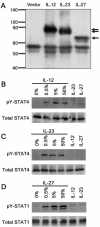
Expression and activity of the scIL-12, scIL-23, and scIL-27 fusion proteins. (A) p3XFLAG-IL-12 (IL-12), p3XFLAG-IL-23 (IL-23), p3XFLAG-IL-27 (IL-27), and p3XFLAG (vector) were transiently transfected into 293T cells. Proteins secreted in the supernatants were immunoprecipitated with the anti-FLAG antibody. The immunoprecipitated proteins were separated on by SDS-12% polyacrylamide gel electrophoresis and subjected to Western blotting analysis with the anti-FLAG antibody. The positions of protein molecular mass markers (in kilodaltons) are shown in the figure, and the three arrows indicate the bands of the scIL-12, scIL-23, and scIL-27 fusion proteins. (B to D) STAT tyrosine phosphorylation assay. 293T cells were transiently transfected with either pME18S-IL-12Rβ1 plus pME18S-IL-12Rβ1 (B), pME18S-IL-12Rβ1 plus p3XFLAG-IL-23R (C), or p3XFLAG-WSX-1/TCCR plus p3XFLAG-gp130 (D). After 36 h, the cells were stimulated for 45 min with the culture supernatant containing either scIL-12 (IL-12) (B), scIL-23 (IL-23) (C), or scIL-27 (IL-27) (D) at final concentrations of 0, 0.5, 5, and 50%. As negative controls, the cells were stimulated for 45 min with the culture supernatant containing either scIL-23/scIL-27 (B), scIL-12/scIL-27 (C), or scIL-12/scIL-23 (D) at a final concentration of 50%. The cells were then subjected to Western blotting with anti-STAT1 (Total STAT1) (D), anti-STAT4 (Total STAT4) (B and C), anti-pY-STAT1 (pY-STAT1) (D), and anti-pY-STAT4 (pY-STAT4) (B and C) antibodies.
FIG. 3.
FIG. 3.
Enhancement of HCV-specific CTL induction by coadministration of either p3XFLAG-IL-12, p3XFLAG-IL-23, or p3XFLAG-IL-27 in the prime-boost immunization. Mice were immunized as follows: priming and the first boosting with 100 μg of either pCEP4-core (A), pCEP4-NS3 (B), or pCEP4-NS4 (C and D), together with 100 μg of either p3XFLAG-IL-12 (circles), p3XFLAG-IL-23 (triangles), p3XFLAG-IL-27 (squares), or p3XFLAG (diamonds), followed by the second boosting with 5 × 10⁷ PFU of either Adex1SR3ST (A) or Adex1CA3269 (B to D). The interval between immunizations was 2 weeks. At 2 to 3 weeks after the last immunization, spleen cells were prepared and stimulated in vitro with syngeneic spleen cells pulsed with 10 μM concentrations of core-132 (A), NS3-1073 (B), NS4-1666 (C), or NS4-1769 (D). After 1 week, 51Cr release assays were performed at various E:T ratios with RMA-HHD cells pulsed with (open symbols) or without (solid symbols) 10 μM concentrations of core-132 (A), NS3-1073 (B), NS4-1666 (C), or NS4-1769 (D) as targets. The data are representative of one of three independent experiments and are shown as the mean ± the standard deviation (SD) of triplicate wells. Similar results were obtained in the three independent experiments.
FIG. 4.
FIG. 4.
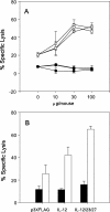
(A) Dose-response curves for the adjuvant plasmids p3XFLAG-IL-12, -IL-23, and -IL-27. Mice were primed and boosted with 100 μg of pCEP4-core, together with various doses (0, 10, 30, and 100 μg) of either p3XFLAG-IL-12 (circles), p3XFLAG-IL-23 (inverted triangles), or p3XFLAG-IL-27 (squares), followed by the second boosting with 5 × 10⁷ PFU of Adex1SR3ST. (B) Synergistic effect of the combination of p3XFLAG-IL-12, -IL-23, and -IL-27. Mice were primed and boosted with 100 μg of pCEP4-core, together with 100 μg of either p3XFLAG, p3XFLAG-IL-12 (IL-12), or the combination of p3XFLAG-IL-12, -IL-23, and -IL-27 (33 μg each) (IL-12/23/27), followed by a second boosting with 5 × 10⁷ PFU of Adex1SR3ST. At 2 to 3 weeks after the last immunization of either the experiment A or B, spleen cells were prepared and stimulated in vitro with syngeneic spleen cells pulsed with 10 μM concentrations of core-132. After 1 week, 51Cr release assays were performed at an E:T ratio of 50 with RMA-HHD cells pulsed with (open symbols or bars) or without (solid symbols or bars) 10 μM core-132 as targets. The data are shown as the mean ± the SD of triplicate wells.
FIG. 5.
FIG. 5.
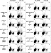
Intracellular IFN-γ staining of HCV-specific CD8+ cells in spleen cells of mice that received coinoculation of either p3XFLAG-IL-12, p3XFLAG-IL-23, or p3XFLAG-IL-27 in the prime-boost immunization. HHD mice were immunized as follows: priming and first boosting with 100 μg of either pCEP4-core (C to J) or pCEP4-NS3 (M to T), along with 100 μg of either p3XFLAG (C, D, M, and N), p3XFLAG-IL-12 (E, F, O, and P), p3XFLAG-IL-23 (G, H, Q, and R), or p3XFLAG-IL-27 (I, J, S, and T); followed by second boosting with 5 × 10⁷ PFU Adex1SR3ST (C to J) or Adex1CA3269 (M to T). Nonimmunized mice (naive) (A, B, K, and L) were used as a negative control. At 2 to 3 weeks after the last immunization, spleen cells were prepared and cultured for 5 h in the presence or absence (A, C, E, G, I, K, M, O, Q, and S) of 10 μM core-132 (B, D, F, H, and J), or 50 μM NS3-1073 (L, N, P, R, and T). After stimulation, cells were stained for their surface expression of CD8 (x axis) and their intracellular expression of IFN-γ (y axis). The numbers shown indicate the percentages of CD8+ cells that are positive for intracellular IFN-γ. The data shown are representative of three independent experiments. Three to five mice per group were used in each experiment, and the spleen cells of the mice per group were pooled.
FIG. 6.
FIG. 6.
Intracellular IFN-γ staining of HCV core-specific CD8+ cells in spleen cells of mice that received preinjections with either p3XFLAG-IL-12, p3XFLAG-IL-23, or p3XFLAG-IL-27, followed by immunization with Adex1SR3ST. HHD mice were primed and boosted with 100 μg of either pCEP4-core (A to H) or pCEP4 (K to P), together with 100 μg of either p3XFLAG (A and B), p3XFLAG-IL-12 (C, D, K, and L), p3XFLAG-IL-23 (E, F, M, and N), or p3XFLAG-IL-27 (G, H, O, and P), followed by the second boosting with 5 × 10⁷ PFU Adex1SR3ST (A to H and K to P). At 2 to 3 weeks after the last immunization, spleen cells were prepared and cultured for 5 h in the presence (B, D, F, H, J, L, N, and P) or absence (A, C, E, G, I, K, M, and O) of 10 μM core-132. After stimulation, cells were stained for their surface expression of CD8 (x axis) and their intracellular expression of IFN-γ (y axis). The numbers shown indicate the percentages of CD8+ cells that are positive for intracellular IFN-γ. The data shown are representative of three independent experiments. We analyzed all data of the three experiments and concluded that the difference between panel B and either panel L (P = 0.005), N (P = 0.038), or P (P = 0.003) was statistically significant as determined by applying the Student t test. At least three mice per group were used in each experiment, and spleen cells of mice per group were pooled.
FIG. 7.
FIG. 7.
Enhancement of CTL induction specific for HCV-core by preinjections of either p3XFLAG-IL-12, p3XFLAG-IL-23, or p3XFLAG-IL-27, followed by immunization with Adex1SR3ST. Mice were preinjected twice with either p3XFLAG-IL-12 (circles), p3XFLAG-IL-23 (inverted triangles), or p3XFLAG-IL-27 (squares), along with pCEP4, followed by immunization with Adex1SR3ST. As a control, mice were primed and boosted with pCEP4-core, together with p3XFLAG, followed by second boosting with Adex1SR3ST (diamonds). The injection doses were 100 μg of plasmid DNA and 5 × 10⁷ PFU of virus, and the interval between immunizations was 2 weeks. After the last immunization, spleen cells were prepared and stimulated in vitro with syngeneic spleen cells pulsed with 10 μM core-132. After 1 week, 51Cr release assays were performed at various E:T ratios with RMA-HHD cells pulsed with (open symbols) or without (solid symbols) 10 μM core-132 as targets. The data are representative of one of three independent experiments and are shown as the mean ± the SD of triplicate wells. Similar results were obtained in the three independent experiments.
FIG. 8.
FIG. 8.
Intracellular IFN-γ staining of HCV core-specific CD8+ cells in spleen cells of mice that received preinjections with either p3XFLAG-IL-12, p3XFLAG-IL-23, or p3XFLAG-IL-27, followed by immunization with either recombinant adenovirus or plasmid DNA. HHD mice were injected with various combinations of 100 μg of plasmid DNA and/or 5 × 10⁷ PFU of recombinant adenovirus as follows: priming and the first boosting with pCEP4-core, followed by the second boosting with Adex1SR3ST (C and D); preinjections twice with either p3XFLAG (E, F, S, and T), p3XFLAG-IL-12 (G, H, M, and N), p3XFLAG-IL-23 (I, J, O, and P), or p3XFLAG-IL-27 (K, L, Q, R, and U-X), followed by immunization with Adex1SR3ST (C-L), Adex1w (M-R), pCEP4-core (S-V), or pCEP4 (W and X). At 2 to 3 weeks after the last immunization, spleen cells were prepared and cultured for 5 h in the presence (B, D, F, H, J, L, N, P, R, T, V, and X) or absence (A, C, E, G, I, K, M, O, Q, S, U, and W) of 10 μM core-132. After in vitro stimulation, the cells were stained for their surface expression of CD8 (x axis) and their intracellular expression of IFN-γ (y axis). The numbers shown indicate the percentages of CD8+ cells that are positive for intracellular IFN-γ. The data shown are representative of three independent experiments. We analyzed all data of the three experiments and concluded that the difference between panel D and either panel H (P = 0.002), J (P = 0.015), or L (P = 0.001) was statistically significant as determined from the Student t test. At least three mice per group were used in each experiment, and spleen cells of mice per group were pooled.

Similar articles

Cited by

References

    1. Alter, M. J., H. S. Margolis, K. Krawczynski, F. N. Judson, A. Mares, W. J. Alexander, P. Y. Hu, J. K. Miller, M. A. Gerber, and R. E. Sampliner. 1992. The natural history of community-acquired hepatitis C in the United States: the Sentinel Counties chronic non-A, non-B hepatitis study team. N. Engl. J. Med. 327:1899-1905. - PubMed
    1. Amara, R. R., F. Villinger, J. D. Altman, S. L. Lydy, S. P. O'Neil, S. I. Staprans, D. C. Montefiori, Y. Xu, J. G. Herndon, L. S. Wyatt, M. A. Candido, N. L. Kozyr, P. L. Earl, J. M. Smith, H. L. Ma, B. D. Grimm, M. L. Hulsey, J. Miller, H. M. McClure, J. M. McNicholl, B. Moss, and H. L. Robinson. 2001. Control of a mucosal challenge and prevention of AIDS by a multiprotein DNA/MVA vaccine. Science 292:69-74. - PubMed
    1. Bain, C., A. Fatmi, F. Zoulin, J.-P. Zarski, C. Trepo, and G. Inchauspe. 2001. Impaired allostimulatory function of dendritic cells in chronic hepatitis C infection. Gastroenterology 120:512-524. - PubMed
    1. Battegay, M., J. Fikes, A. M. Di Bisceglie, P. A. Wentworth, A. Sette, E. Celis, W.-M. Ching, A. Grakoui, C. M. Rice, K. Kurokohchi, J. A. Berzofsky, J. H. Hoofnagle, S. M. Feinstone, and T. Akatsuka. 1995. Patients with chronic hepatitis C have circulating cytotoxic T cells which recognize hepatitis C virus-encoded peptides binding to HLA-A2.1 molecules. J. Virol. 69:2462-2470. - PMC - PubMed
    1. Belladonna, M. L., J.-C. Renauld, R. Bianchi, C. Vacca, F. Fallarino, C. Orabona, M. C. Fioretti, U. Grohmann, and P. Puccetti. 2001. IL-23 and IL-12 have overlapping, but distinct, effects on murine dendritic cells. J. Immunol. 168:5448-5454. - PubMed

Publication types

MeSH terms