. 2004 Sep;78(17):9132-43.
doi: 10.1128/JVI.78.17.9132-9143.2004.

Factors that increase the effective concentration of CXCR4 dictate feline immunodeficiency virus tropism and kinetics of replication

Affiliations

Factors that increase the effective concentration of CXCR4 dictate feline immunodeficiency virus tropism and kinetics of replication

Aymeric de Parseval et al. J Virol. 2004 Sep.

Abstract

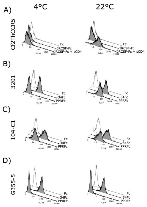
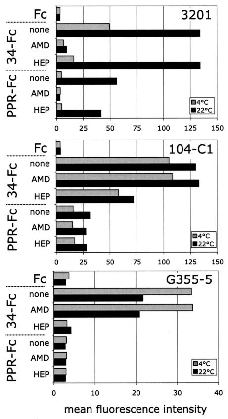
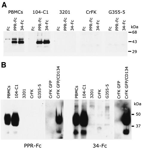
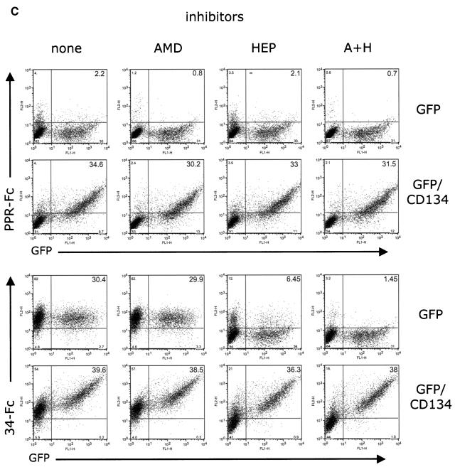
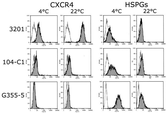
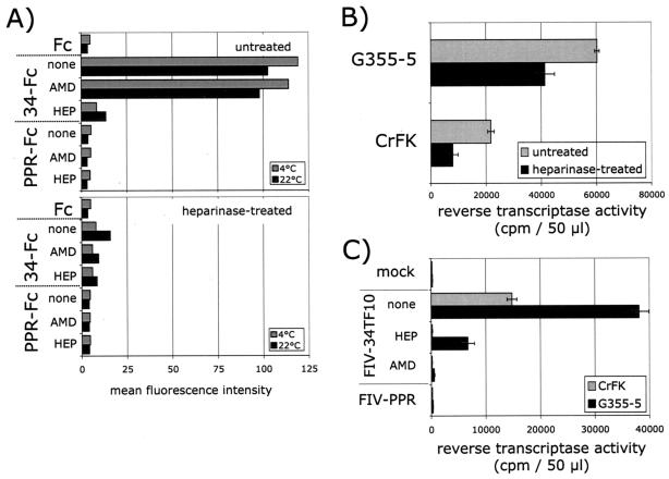
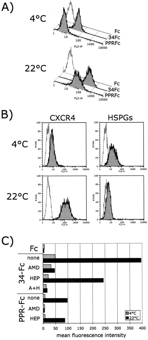
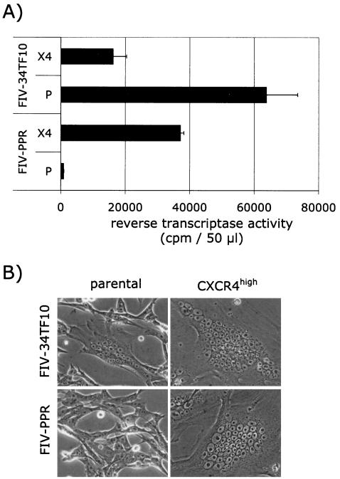
The surface glycoprotein (gp95) of the feline immunodeficiency virus (FIV) binds in a strain-specific manner to several cell surface molecules, including CXCR4, heparan sulfate proteoglycans (HSPGs), DC-SIGN, and a 43-kDa cell surface receptor on T cells recently identified as CD134 by M. Shimojima et al. (Science 303:1192-1195, 2004). CXCR4 is the entry receptor in all known cases, and the other molecules act as binding receptors to help facilitate infection. In this report, we confirm and extend the findings regarding CD134 as a primary receptor for FIV. In addition, we show that temperature critically influences the binding properties of FIV gp95 to CXCR4 and HSPGs. The data show that gp95 of the field strain FIV-PPR bound to CXCR4 at 22 degrees C, whereas binding was not detected at 4 degrees C. In contrast, binding of the laboratory adapted FIV-34TF10 gp95 was observed at either 4 degrees C or 22 degrees C, albeit at increased levels at the higher temperature. The level of CXCR4 increased after the temperature was switched from 4 to 22 degrees C, whereas the level of HSPGs decreased, resulting in higher binding of gp95 from both strains to CXCR4 and lower binding of gp95 of FIV-34TF10 to HSPGs (FIV-PPR gp95 does not bind to these molecules). The findings also show that HSPGs facilitate the CXCR4-mediated infectivity of CrFK and G355-5 cells by FIV-34TF10. These two nonlymphoid cell lines express very low levels of CXCR4 and are permissive to FIV-34TF10 but not to productive infection by FIV-PPR. However, overexpression of human CXCR4 in CrFK or G-355-5 cells resulted in extensive cell fusion and infection by FIV-PPR. Taken together, these findings indicate that factors that increase the effective concentration of CXCR4 enhance FIV infectivity and may involve (i) temperature or ligand-induced conformational changes in CXCR4 that enhance SU binding, (ii) coreceptor interactions with gp95 that either alter gp95 conformation to enhance CXCR4 binding and/or raise the localized concentration of receptor or ligand, or (iii) direct increase in CXCR4 concentration via overexpression.


Figures

FIG. 1.
FIG. 1.
Binding profile of FIV SU adhesins: conformation-dependent binding of FIV SU to CXCR4. (A) FACS analysis of the binding of HIV-1 JRCSF-Fc to the CD4 CCR5+ Cf2Th synCCR5 cells requires sCD4 and a temperature shift from 4 to 22°C. (B) FACS analysis of the binding of FIV PPR-Fc and 34-Fc to the CXCR4+ 3201 feline lymphoma cells. Binding of PPR-Fc to CXCR4 requires only a shift in temperature; likewise, binding of 34-Fc is increased after the temperature shift. (C and D) FACS analysis of the binding of PPR-Fc and 34-Fc at 4 and 22°C on 104-C1 and G355-5, respectively. Fc alone was used as a negative control.
FIG. 2.
FIG. 2.
Inhibition profile of FIV SU adhesins. Inhibition of PPR-Fc and 34-Fc binding to 3201, 104-C1, and G355-5 cells at 4 and 22°C by AMD3100 and heparin. Cells were incubated with the indicated SU adhesins in the absence (none) or presence of AMD3100 (AMD), heparin (HEP), or both (A+H). SU binding, expressed as the mean fluorescence intensity, was analyzed by FACS analysis as described in Materials and Methods. Heparin and AMD3100 were used at 1 μg/ml. Fc alone was used as a negative control.
FIG. 3.
FIG. 3.
A 43-kDa binding receptor for gp95 on PBMC and the IL-2-dependent T-cell line 104-C1 corresponds to the T-cell marker, CD134. (A) Immunoprecipitation studies identified a 43-kDa protein species on PBMC and 104-C1 cells that specifically interacts with FIV SU adhesins. Biotinylated cell lysates were incubated with the indicated adhesins. Complexes were resolved by SDS-PAGE, and gp95-binding proteins were revealed by Western blotting with a neutravidin-HRP antibody. This receptor was not immunoprecipitated from 3201, CrFK, or G355-5 cells. (B) A modified virus overlay assay shows that FIV SU adhesins interact with a 43-kDa protein species on PBMC and 104-C1 cells but not 3201, CrFK, and G355-5 cells. A similarly sized molecule was bound by both PPR and 34TF10 SU adhesins on CrFK transduced with MIGR1-GFP/CD134 (CrFK GFP/CD134) but not on CrFK transduced with MIGR1 vector expressing only GFP (CrFK GFP). The indicated cell lysates were resolved by SDS-PAGE. Blots were then overlaid with the indicated adhesins. Binding of gp95 was revealed by an anti-Fc-HRP antibody. (C) FACS analysis revealing the specific interaction of FIV SU with CD134. CrFK cells were transduced with MIGR1-GFP or MIGR1-GFP/CD134. Binding with PPR-Fc and 34-Fc was assessed by FACS analysis in the absence (none) or presence of AMD3100 (AMD), heparin (HEP), or both (H+P). Note that neither AMD3100, heparin, or a combination of both inhibitors inhibited the binding of either SU adhesin to CrFK expressing CD134. Heparin and AMD3100 were used at 1 μg/ml.
FIG. 3.
FIG. 3.
A 43-kDa binding receptor for gp95 on PBMC and the IL-2-dependent T-cell line 104-C1 corresponds to the T-cell marker, CD134. (A) Immunoprecipitation studies identified a 43-kDa protein species on PBMC and 104-C1 cells that specifically interacts with FIV SU adhesins. Biotinylated cell lysates were incubated with the indicated adhesins. Complexes were resolved by SDS-PAGE, and gp95-binding proteins were revealed by Western blotting with a neutravidin-HRP antibody. This receptor was not immunoprecipitated from 3201, CrFK, or G355-5 cells. (B) A modified virus overlay assay shows that FIV SU adhesins interact with a 43-kDa protein species on PBMC and 104-C1 cells but not 3201, CrFK, and G355-5 cells. A similarly sized molecule was bound by both PPR and 34TF10 SU adhesins on CrFK transduced with MIGR1-GFP/CD134 (CrFK GFP/CD134) but not on CrFK transduced with MIGR1 vector expressing only GFP (CrFK GFP). The indicated cell lysates were resolved by SDS-PAGE. Blots were then overlaid with the indicated adhesins. Binding of gp95 was revealed by an anti-Fc-HRP antibody. (C) FACS analysis revealing the specific interaction of FIV SU with CD134. CrFK cells were transduced with MIGR1-GFP or MIGR1-GFP/CD134. Binding with PPR-Fc and 34-Fc was assessed by FACS analysis in the absence (none) or presence of AMD3100 (AMD), heparin (HEP), or both (H+P). Note that neither AMD3100, heparin, or a combination of both inhibitors inhibited the binding of either SU adhesin to CrFK expressing CD134. Heparin and AMD3100 were used at 1 μg/ml.
FIG. 4.
FIG. 4.
Temperature-dependent detection of CXCR4 and HSPGs. Cells were labeled at the indicated temperatures, and the detection of CXCR4 and HSPGs was carried out by FACS analysis. The switch from 4 to 22°C induced an increase in CXCR4 detection that was most noticeable on 3201 cells. In contrast, HSPG detection was higher at 4°C and markedly decreased when the temperature was raised to 22°C.
FIG. 5.
FIG. 5.
HSPGs facilitate TCA FIV binding and infection of G355-5 and CRFK cells. (A) Binding of FIV SU adhesins was analyzed at 4 and 22°C before and after heparinase treatment of G355-5 cells. Heparinase treatment significantly reduced the binding of 34-Fc. Inhibition of binding by AMD3100 (AMD) and heparin (HEP) showed that residual binding after heparinase treatment is mediated by both CXCR4 and HSPGs. (B) Heparinase treatment reduced FIV-34TF10 infectivity. CrFK and G355-5 Cells were treated in the absence or presence of heparinase, washed, and then infected with FIV-34TF10. Virus production was monitored by an RT assay at 7 days postinfection. (C) Heparin efficiently neutralized FIV infection. Cells were mock infected or infected with FIV-34TF10 and FIV-PPR. Cells infected with FIV-34TF10 were treated in the absence (none) or presence of heparin (10 μg/ml, HEP) or AMD3100 (1 μg/ml, AMD). Virus production was monitored by an RT assay at 7 days postinfection.
FIG. 6.
FIG. 6.
Overexpression of CXCR4 results in efficient binding of PPR-Fc on G355-5 cells. (A) Binding of FIV SU adhesins at 4 and 22°C on CXCR4high G355-5 cells. At 22°C, PPR-Fc efficiently bound the CXCR4high cells, whereas no binding was observed at 4°C. (B) Detection profile of CXCR4 and HSPGs at 4 and 22°C on CXCR4high G355-5 cells. As with the 3201 cells (Fig. 4), CXCR4 detection was temperature dependent and increased after the temperature was raised to 22°C. The detection profile of HSPGs was similar to parental cells (Fig. 4). (C) Inhibition of binding of FIV SU adhesins to CXCR4high cells. Cells were incubated at 4 or 22°C with the indicated SU adhesins in the absence (none) or presence of AMD3100 (AMD), heparin (HEP), or both (A+H). SU binding, expressed as the mean fluorescence intensity, was analyzed by FACS analysis as described in Materials and Methods. Heparin and AMD3100 were used at 1 μg/ml. Fc alone was used as a negative control.
FIG. 7.
FIG. 7.
Overexpression of CXCR4 renders G355-5 cells permissive to FIV-PPR infection. (A) Parental (P) and CXCR4high (X4) G355-5 cells were infected with FIV-34TF10 and FIV-PPR, and virus production was monitored at 5 days postinfection. (B) Massive cell fusion was observed in the CXCR4high cell population infected with either FIV strain at 3 days postinfection. Small syncytia with fewer nuclei were observed only with the parental cells infected with FIV-34TF10.
FIG. 8.
FIG. 8.
The block in infection of 3201 cells occurs at a postbinding level. (A) FACS analysis comparing binding of PPR-Fc at 22°C to 3201 (not permissive) and 104-C1 (permissive) cells. (B) PCR-based assay showing an apparent block of replication in 3201 cells. 3201 (lanes 1 and 2) and 104-C1 (lanes 3 and 4) cells were mock infected (lanes 1 and 3) or FIV-PPR infected (lanes 2 and 4), and newly synthesized viral DNA and viral RNA were analyzed at 20 and 72 h postinfection, respectively. PCR with primer pairs specific for a CD4 intron and β-actin were used as controls of DNA and RNA extractions, respectively.

Similar articles

Cited by

References

    1. Babcock, G. J., T. Mirzabekov, W. Wojtowicz, and J. Sodroski. 2001. Ligand binding characteristics of CXCR4 incorporated into paramagnetic proteoliposomes. J. Biol. Chem. 276:38433-38440. - PubMed
    1. Baribaud, F., T. G. Edwards, M. Sharron, A. Brelot, N. Heveker, K. Price, F. Mortari, M. Alizon, M. Tsang, and R. W. Doms. 2001. Antigenically distinct conformations of CXCR4. J. Virol. 75:8957-8967. - PMC - PubMed
    1. Beebe, A. M., N. Dua, T. G. Faith, P. F. Moore, N. C. Pedersen, and S. Dandekar. 1994. Primary stage of feline immunodeficiency virus infection: viral dissemination and cellular targets. J. Virol. 68:3080-3091. - PMC - PubMed
    1. Bobardt, M. D., A. C. Saphire, H. C. Hung, X. Yu, B. Van der Schueren, Z. Zhang, G. David, and P. A. Gallay. 2003. Syndecan captures, protects, and transmits HIV to T lymphocytes. Immunity 18:27-39. - PubMed
    1. Brelot, A., N. Heveker, K. Adema, M. J. Hosie, B. Willett, and M. Alizon. 1999. Effect of mutations in the second extracellular loop of CXCR4 on its utilization by human and feline immunodeficiency viruses. J. Virol. 73:2576-2586. - PMC - PubMed

Publication types

MeSH terms

LinkOut - more resources