. 2004 Sep;78(17):9144-53.
doi: 10.1128/JVI.78.17.9144-9153.2004.

The PA subunit is required for efficient nuclear accumulation of the PB1 subunit of the influenza A virus RNA polymerase complex

Affiliations

The PA subunit is required for efficient nuclear accumulation of the PB1 subunit of the influenza A virus RNA polymerase complex

Ervin Fodor et al. J Virol. 2004 Sep.

Abstract

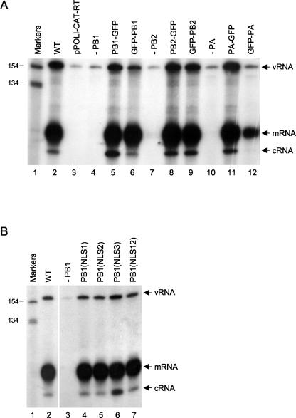
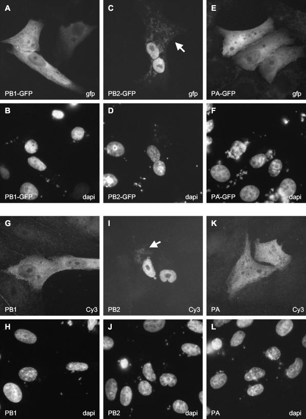
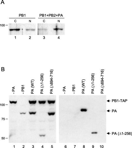
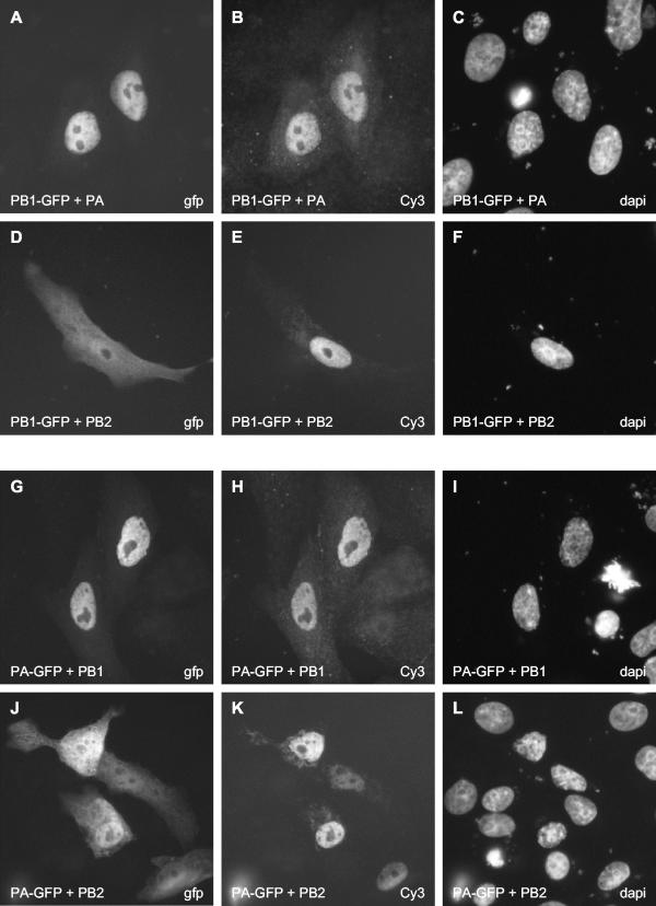
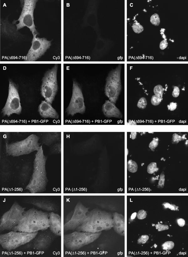
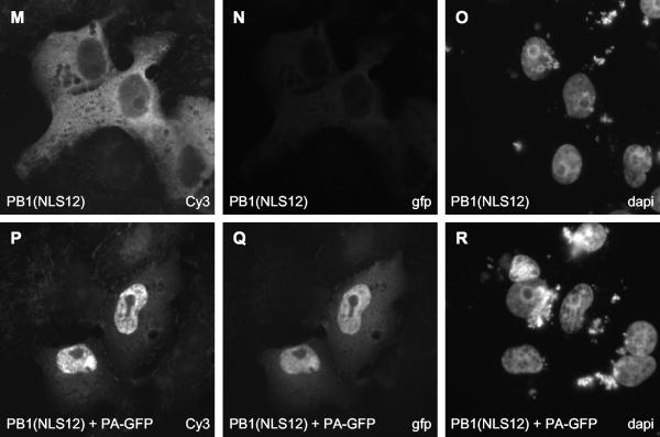
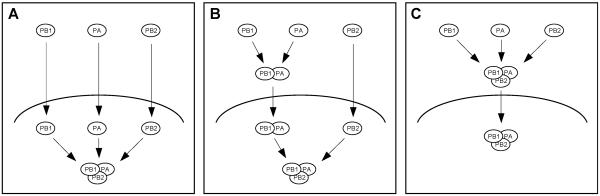
The RNA genome of influenza virus is transcribed and replicated by the viral RNA polymerase complex in the cell nucleus. We have generated green fluorescent protein (GFP)-tagged polymerase subunits to study the assembly of the polymerase complex. Our results show that individually expressed polymerase basic protein 1 (PB1) and polymerase acidic protein (PA) subunits were distributed in both the cytoplasm and the nucleus, while the polymerase basic protein 2 (PB2) subunit accumulated in the nucleus. Although it has been reported that PB1 alone accumulates in the nucleus, we demonstrate that PB1 requires the coexpression of PA for efficient nuclear accumulation. Our results support a model which proposes that PB1 and PA are transported into the nucleus as a complex.


Figures

FIG. 1.
FIG. 1.
Primer extension assays of vRNA, mRNA, and cRNA. (A) Effects of GFP tags on transcription and replication of influenza virus RNA. Primer extension assays of vRNA, mRNA, and cRNA isolated from 293T cells expressing influenza virus polymerase proteins (not tagged, i.e., wild type [WT]; tagged with GFP at the C terminus [PB1-GFP, PB2-GFP, and PA-GFP]; or tagged with GFP at the N terminus [GFP-PB1, GFP-PB2, and GFP-PA]), NP, and vRNA-like CAT RNA were performed as described previously (12). Negative controls include the analysis of RNA isolated from cells transfected with pPOLI-CAT-RT alone (lane 3) or with the omission of pcDNA-PB1 (lane 4), pcDNA-PB2 (lane 7), or pcDNA-PA (lane 10). (B) Effects of mutations in the potential NLSs of PB1 on the transcription and replication of influenza virus RNA. Primer extension assays of vRNA, mRNA, and cRNA isolated from 293T cells expressing influenza virus PB1 (wild type [WT] or the indicated mutants), PB2, PA, NP, and vRNA-like CAT RNA were performed as described previously (12). Positions of vRNA, mRNA, and cRNA signals are indicated on the right. Sizes of markers (32P-labeled 1-kb ladder [Invitrogen]) are indicated on the left.
FIG. 2.
FIG. 2.
Intracellular distribution of GFP-tagged and nontagged polymerase subunits in transfected Vero cells. Cells expressing GFP-tagged polymerase subunits (A, C, and E) were analyzed directly for GFP expression by fluorescence microscopy. Cells expressing nontagged polymerase subunits (G, I, and K) were examined after staining with the corresponding subunit-specific primary and Cy3-conjugated secondary antibodies as described in Materials and Methods. Panels B, D, F, H, J, and L show the same fields of cells as panels A, C, E, G, I, and K, respectively, stained for DNA to indicate the locations of nuclei. The expressed protein is indicated in the bottom left corner of each panel. The method of detection (GFP, Cy3, or DAPI) is indicated in the bottom right corner of each panel. The white arrows in panel C and I indicate the mitochondrial localizations of PB2-GFP and PB2, respectively.
FIG. 3.
FIG. 3.
(A) Intracellular distribution of PB1 in the absence or presence of PB2 and PA. 293T cells expressing PB1 (lanes 1 and 2) or PB1, PB2, and PA (lanes 3 and 4) were fractionated into cytoplasmic (C) and nuclear (N) fractions, and equivalent amounts were subjected to SDS-PAGE and Western blotting with a polyclonal anti-PB1 antibody. (B) Expression and binding to PB1 of mutant PA proteins. Extracts from 293T cells expressing PA (wild type [WT] or the indicated deletion mutants) and TAP-tagged PB1 (37) were analyzed by SDS-PAGE and Western blotting with a polyclonal anti-PA antibody before (left panel) and after (right panel) purification. Note the cross-reaction of PB1-TAP in the left panel due to the presence of the TAP tag. Sizes (in kilodaltons) are indicated on the left.
FIG. 4.
FIG. 4.
Effect of PA or PB2 on the localization of PB1-GFP and effect of PB1 or PB2 on the localization of PA-GFP in transfected Vero cells. Cells expressing the indicated combinations of proteins (see bottom left of each panel) were analyzed directly for the GFP-tagged polymerase subunits (A, D, G, and J) and stained for the nontagged subunits with the corresponding subunit-specific primary and Cy3-conjugated secondary antibodies (B, E, H, and K). The three panels in each row show the same field of cells. The method of detection (GFP, Cy3, or DAPI) is indicated in the bottom right corner of each panel.
FIG. 5.
FIG. 5.
Effect of mutations in PA and PB1 on the nuclear accumulation of PB1. PA deletion mutants (A, D, G, and J) and the PB1 NLS12 mutant (M and P) were detected with rabbit polyclonal anti-PA and anti-PB1 antibodies, respectively, followed by staining with a Cy3-conjugated anti-rabbit secondary antibody. PB1-GFP (E and K) and PA-GFP (Q) were detected directly by fluorescence microscopy. The three panels in each row show the same field of cells. The method of detection (GFP, Cy3, or DAPI) is indicated in the bottom right corner of each panel.
FIG. 5.
FIG. 5.
Effect of mutations in PA and PB1 on the nuclear accumulation of PB1. PA deletion mutants (A, D, G, and J) and the PB1 NLS12 mutant (M and P) were detected with rabbit polyclonal anti-PA and anti-PB1 antibodies, respectively, followed by staining with a Cy3-conjugated anti-rabbit secondary antibody. PB1-GFP (E and K) and PA-GFP (Q) were detected directly by fluorescence microscopy. The three panels in each row show the same field of cells. The method of detection (GFP, Cy3, or DAPI) is indicated in the bottom right corner of each panel.
FIG. 6.
FIG. 6.
Models for the transport and assembly of the influenza virus RNA polymerase. (A) Subunits PB1, PB2, and PA are transported into the nucleus individually and form a complex in the nucleus. (B) PB1 and PA interact in the cytoplasm and are transported into the nucleus as a dimer. PB2 enters the nucleus as a monomer and binds to PB1-PA in the nucleus. (C) PB1, PB2, and PA interact in the cytoplasm and are transported into the nucleus as a trimeric complex.

References

    1. Akkina, R. K., T. M. Chambers, D. R. Londo, and D. P. Nayak. 1987. Intracellular localization of the viral polymerase proteins in cells infected with influenza virus and cells expressing PB1 protein from cloned cDNA. J. Virol. 61:2217-2224. - PMC - PubMed
    1. Area, E., J. Martín-Benito, P. Gastaminza, E. Torreira, J. M. Valpuesta, J. L. Carrascosa, and J. Ortín. 2004. 3D structure of the influenza virus polymerase complex: localization of subunit domains. Proc. Natl. Acad. Sci. USA 101:308-313. - PMC - PubMed
    1. Argos, P. 1988. A sequence motif in many polymerases. Nucleic Acids Res. 16:9909-9916. - PMC - PubMed
    1. Bárcena, J., M. Ochoa, S. de la Luna, J. A. Melero, A. Nieto, J. Ortín, and A. Portela. 1994. Monoclonal antibodies against influenza virus PB2 and NP polypeptides interfere with the initiation step of viral mRNA synthesis in vitro. J. Virol. 68:6900-6909. - PMC - PubMed
    1. Biswas, S. K., P. L. Boutz, and D. P. Nayak. 1998. Influenza virus nucleoprotein interacts with influenza virus polymerase proteins. J. Virol. 72:5493-5501. - PMC - PubMed

Publication types

MeSH terms