. 2004 Sep;78(17):9446-57.
doi: 10.1128/JVI.78.17.9446-9457.2004.

Kinetic analysis of the interactions between vaccinia virus complement control protein and human complement proteins C3b and C4b

Affiliations

Kinetic analysis of the interactions between vaccinia virus complement control protein and human complement proteins C3b and C4b

John Bernet et al. J Virol. 2004 Sep.

Abstract

The vaccinia virus complement control protein (VCP) is an immune evasion protein of vaccinia virus. Previously, VCP has been shown to bind and support inactivation of host complement proteins C3b and C4b and to protect the vaccinia virions from antibody-dependent complement-enhanced neutralization. However, the molecular mechanisms involved in the interaction of VCP with its target proteins C3b and C4b have not yet been elucidated. We have utilized surface plasmon resonance technology to study the interaction of VCP with C3b and C4b. We measured the kinetics of binding of the viral protein to its target proteins and compared it with human complement regulators factor H and sCR1, assessed the influence of immobilization of ligand on the binding kinetics, examined the effect of ionic contacts on these interactions, and sublocalized the binding site on C3b and C4b. Our results indicate that (i) the orientation of the ligand is important for accurate determination of the binding constants, as well as the mechanism of binding; (ii) in contrast to factor H and sCR1, the binding of VCP to C3b and C4b follows a simple 1:1 binding model and does not involve multiple-site interactions as predicted earlier; (iii) VCP has a 4.6-fold higher affinity for C4b than that for C3b, which is also reflected in its factor I cofactor activity; (iv) ionic interactions are important for VCP-C3b and VCP-C4b complex formation; (v) VCP does not bind simultaneously to C3b and C4b; and (vi) the binding site of VCP on C3b and C4b is located in the C3dg and C4c regions, respectively.


Figures

FIG. 1.
FIG. 1.
Schematic representation of structures of VCP, factor H, and sCR1. VCP is entirely composed of four SCR or CCP modules, whereas the human complement regulators factor H and complement receptor 1 are composed of 20 and 30 SCRs, respectively. The SCR domains of each protein are numbered, and the binding domains for C3b and C4b are identified (3, 19, 20, 26, 41, 48, 50). The relative binding activity of VCP to that of factor H and sCR1 is shown on the right. These activities are based on the data presented in Table 1. formula image, Factor H binds to C4b only in a buffer with very low ionic strength (47).
FIG. 2.
FIG. 2.
Analysis of the binding of immobilized VCP to C3b and C4b by SPR. The left panels show sensogram overlays for interactions between immobilized VCP and C3b or C4b. The concentration of analyte injected is indicated at the right of each sensogram overlay. Solid lines correspond to the global fitting of the data simultaneously. Both C3b and C4b data fit to a bivalent analyte model (A+B ↔ AB; AB + B ↔ AB2; BIAevaluation 3.2). The right panels show linear transformations of the association phase data for the respective sensogram data shown on the left. The straight lines are linear least square fits to the data.
FIG. 3.
FIG. 3.
Site-specific biotinylation of C3b and C4b. The free −SH groups of C3b and C4b were labeled with PEO-maleimide biotin, and labeled proteins were cleaved into iC3b or C3c and C3dg and C4c and C4d and analyzed by SDS-PAGE and Western blotting as described in Materials and Methods. C3b cleavage in the presence of factors H and I results in cleavage of the α′ chain into N-terminal 68-kDa and C-terminal 43-kDa fragments; the appearance of these fragments indicates the generation of iC3b, whereas C3b cleavage in the presence of sCR1 and factor I results in cleavage of the α′ chain into N-terminal 25-kDa fragment, C3dg, and C-terminal 43-kDa fragments, which indicates the generation of C3c and C3dg. C4b cleavage in the presence of sCR1 and factor I results in cleavage of the α′ chain into N-terminal 25-kDa, C-terminal 16-kDa, and central C4d fragments; the appearance of these fragments indicates the generation of C4c and C4d. (A) Diagram showing covalent attachment of C3b and C4b to the activating surface. (B) Diagram depicting the experimental design. Site-specific biotinylated C3b and C4b were immobilized on a streptavidin chip (SA chip). (C) Coomassie blue staining (left) and Western blot (right) of biotinylated C3b and its cleavage products. Lane 1, biotinylated C3b; lane 2, biotinylated C3b cleaved with factors I and H; lane 3, biotinylated C3b cleaved with factor I and sCR1. (D) Schematic representation of C3b, C3c, and C3dg structures. Arrows indicate factor I-mediated cleavages generated in the presence of cofactors and closed balloon indicates the location of −SH group labeled with biotin. (E) Coomassie blue staining (left) and Western blot (right) of biotinylated C4b and its cleavage products. Lane 1, biotinylated C4b; lane 2, biotinylated C4b cleaved with factor I and sCR1. (F) Schematic representation of C4b, C4c, and C4d structures. Arrows indicate factor I-mediated cleavages generated in the presence of sCR1, and the closed balloon indicates the location of −SH group labeled with biotin.
FIG. 4.
FIG. 4.
Analysis of binding of VCP to immobilized C3b and C4b. The left panels show overlay plots of the binding of immobilized C3b and C4b to VCP. Various concentrations of VCP were injected over a streptavidin chip immobilized with C3b or C4b. Solid lines represent the global fitting of the data to a 1:1 Langmuir binding model (A+B ↔ AB; BIAevaluation 3.2). The right panels show linear transformations of the association data for the respective sensogram data shown on the left. The straight lines are linear least-square fits to the data.
FIG. 5.
FIG. 5.
Binding of sCR1 and factor H to immobilized C3b and C4b. The left panels show overlay plots for binding of sCR1, factor H to immobilized C3b, and sCR1 to immobilized C4b. Various concentrations of analytes were injected over streptavidin chips containing biotinylated C3b or C4b. Solid lines shown in the C3b-CR1 panel represent the global fitting of the data to a bivalent analyte model (A+B ↔ AB; AB + B ↔ AB2; BIAevaluation 3.2). The middle panels show the linear transformation of the association phase data of the respective sensogram data shown on the left. The straight lines are linear least-square fits to the data. The inset shows values of ks (determined from the slope of the fits of the association data) replotted against analyte concentration. The slope of this plot provided ka. The right panels show the linear transformation of the highest concentration of the dissociation phase data of the respective analyte shown on the left. The slope of the fits provided the off rates (kd).
FIG. 6.
FIG. 6.
Comparison of factor I cofactor activity of VCP for C3b and C4b. Equimolar concentrations of C3b or C4b and factor I were incubated in 10 mM sodium phosphate (pH 7.4) containing 145 mM NaCl with increasing concentrations of VCP at 37°C for 4 h (upper and middle panels). Cleavage products were visualized by running the samples on 8% and 9% SDS-PAGE gels for C3b and C4b, respectively, and staining with Coomassie blue. The intensities of the α′ chains of C3b and C4b were determined by densitometric analysis and are represented graphically (lower panel).
FIG. 7.
FIG. 7.
Effect of NaCl concentration on the binding of VCP to C3b and C4b. Sensogram overlays for binding of immobilized VCP to C3b (125 nM) and C4b (100 nM) in the presence of various concentrations of NaCl are shown. On the left, the binding response (RU) is plotted against time. On the right, the maximum binding response obtained for each buffer condition is plotted against the NaCl concentration.
FIG. 8.
FIG. 8.
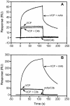
Simultaneous binding of C3b and C4b to VCP. (A) VCP (0.6 μM), VCP (0.6 μM) preincubated with C4b (0.56 μM), or VCP (0.6 μM) preincubated with anti-VCP MAb (0.4 μM [this antibody does not inhibit the functional activity of VCP]) at 25°C for 30 min was injected over a streptavidin chip containing biotinylated C3b, and the association and dissociation phases were monitored. (B) VCP (0.6 μM), VCP (0.6 μM) preincubated with C3b (0.6 μM), or VCP (0.6 μM) preincubated with anti-VCP MAb (0.2 μM) at 25°C for 30 min was injected over a streptavidin chip containing biotinylated C4b, and binding and dissociation were monitored.
FIG. 9.
FIG. 9.
Binding of VCP to immobilized C3b and C4b fragments. The left panels show sensograms for interactions between VCP and C3b or C4b fragments. VCP (1.3 μM) was injected over a streptavidin chip immobilized either with C3b fragments (C3c-biotin and C3dg-biotin) or with C4b fragments (C4c-biotin and C4d-biotin). The right panels show overlay plots for interactions between immobilized C3dg or C4c and VCP. The small arrow in the C3dg-VCP panel indicates the time point used for evaluating steady-state affinity data. Solid lines shown in the C4c-VCP panel represent the global fitting of the data to a 1:1 Langmuir binding model (A + B ↔ AB; BIAevaluation 3.2).

References

    1. Ahearn, J. M., M. B. Fischer, D. Croix, S. Goerg, M. Ma, J. Xia, X. Zhou, R. G. Howard, T. L. Rothstein, and M. C. Carroll. 1996. Disruption of the Cr2 locus results in a reduction in B-1a cells and in an impaired B-cell response to T-dependent antigen. Immunity 4:251-262. - PubMed
    1. Alcami, A., and U. H. Koszinowski. 2000. Viral mechanisms of immune evasion. Trends Microbiol. 8:410-418. - PMC - PubMed
    1. Alsenz, J., J. D. Lambris, T. F. Schulz, and M. P. Dierich. 1984. Localization of the complement-component-C3b-binding site and the cofactor activity for factor I in the 38-kDa tryptic fragment of factor H. Biochem. J. 224:389-398. - PMC - PubMed
    1. Arnaout, M. A., N. Dana, J. Melamed, R. Medicus, and H. R. Colten. 1983. Low ionic strength or chemical cross-linking of monomeric C3b increases its binding affinity to the human complement C3b receptor. Immunology 48:229-237. - PMC - PubMed
    1. Bernet, J., J. Mullick, A. K. Singh, and A. Sahu. 2003. Viral mimicry of the complement system. J. Biosci. 28:249-264. - PMC - PubMed

Publication types

LinkOut - more resources