. 2004 Sep;78(17):9544-51.
doi: 10.1128/JVI.78.17.9544-9551.2004.

Pathogenesis of acute viral disease induced in fish by carp interstitial nephritis and gill necrosis virus

Affiliations

Pathogenesis of acute viral disease induced in fish by carp interstitial nephritis and gill necrosis virus

Eli Pikarsky et al. J Virol. 2004 Sep.

Abstract

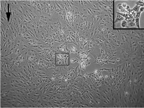
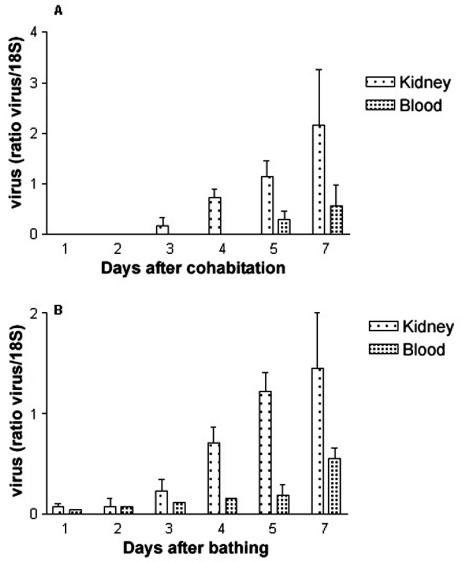
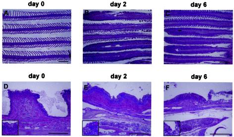
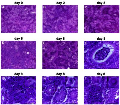
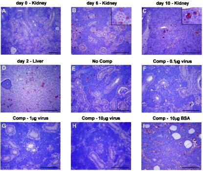
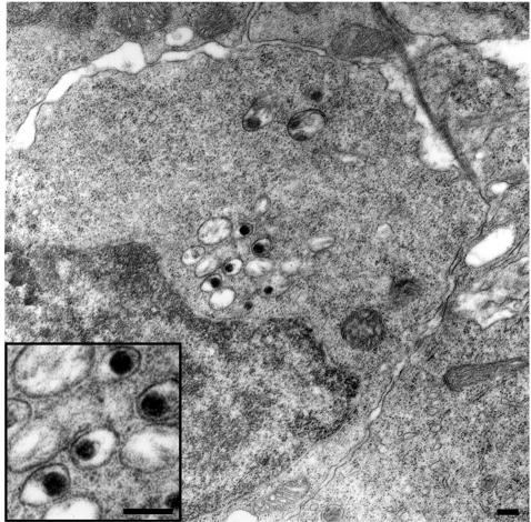
A lethal disease of koi and common carp (species Cyprinus carpio) has afflicted many fish farms worldwide since 1998, causing severe financial losses. Morbidity and mortality are restricted to common carp and koi and appear in spring and autumn, when water temperatures are 18 to 28 degrees C. We have isolated the virus causing the disease from sick fish, propagated it in koi fin cell culture, and shown that virus from a single clone causes lethal disease in carp and koi upon infection. Intraperitoneal virus injection or bathing the fish in virus-containing water kills 85 to 100% of the fish within 7 to 21 days. This virus is similar to the previously reported koi herpesvirus; however, it has characteristics inconsistent with the herpesvirus family, and thus we have called it carp interstitial nephritis and gill necrosis virus. We examined the pathobiology of this disease in carp by using immunohistochemistry and PCR. We found large amounts of the virus in the kidneys of sick fish and smaller amounts in liver and brain. A rapid increase in the viral load in the kidneys was detected by using both immunofluorescence and semiquantitative PCR. Histological analyses of fish at various times after infection revealed signs of interstitial nephritis as early as 2 days postinfection, which increased in severity up to 10 days postinfection. There was severe gill disease evidenced by loss of villi with accompanying inflammation in the gill rakers. Minimal focal inflammation was noted in livers and brains. This report describes the etiology and pathology of a recently described viral agent in fish.


Figures

FIG. 1.
FIG. 1.
Cytopathic effect induced by CNGV. KFCs were infected with CNGV and incubated at a permissive temperature of 22°C. At 4 to 5 days p.i., cytopathic effect can be observed. Cells become enlarged and develop abundant endoplasmic vacuoles (center box; the inset at upper right shows the boxed area at a higher magnification). The arrow indicates uninfected cells.
FIG. 2.
FIG. 2.
Mortality of fish infected with cloned virus. Fish (n = 50; average weight, 100 g) were injected with 0.2 ml of a solution containing approximately 102 PFU of the cloned virus. The control group was composed of fish (n = 50; average weight, 100 g) injected with 0.2 ml of a solution containing medium harvested from uninfected cultures.
FIG. 3.
FIG. 3.
Kinetics of the appearance of viral DNA in infected fish. Semiquantitative PCR analysis of CNGV DNA was performed on DNA extracted from fish blood samples or kidneys following infection by cohabitation (A) or bathing (B). The CNGV DNA levels are normalized against those of 18S ribosomal DNA in the same sample. Each bar represents the mean for five fish ± standard error of the mean.
FIG. 4.
FIG. 4.
Immunofluorescence assay for the presence of CNGV in fish organs. Touch imprints of kidney (A), liver (B), and brain (C) from infected fish and of kidney from healthy fish (D) were incubated with rabbit anti-CNGV serum (1:1,000). Antibodies were detected by using fluorescein isothiocyanate-conjugated swine anti-rabbit antibodies.
FIG. 5.
FIG. 5.
CNGV induces gill inflammation as early as 2 days p.i. Carps were infected with CNGV and harvested at the indicated days p.i. Gills were collected and subjected to histological analyses. (A to C) Gill filaments. Normally gill filaments are slender structures containing numerous lamellae (A). As early as 2 days p.i. (B), many lamellae are infiltrated by inflammatory cells. At 6 days p.i. (C) and onwards, all lamellae are heavily infiltrated. (D to F) Gill rakers. As early as 2 days p.i. (E), an increased inflammatory infiltrate is present in the subepithelial zone. In addition, at the bottom of the photomicrograph a congested vessel in the gill arch is seen. At 6 days p.i. (F), the inflammatory process is more pronounced, with sloughing of the overlying epithelium (upper right). This is accompanied by increased congestion and edema. All of the sections were stained with hematoxylin and eosin. The insets in the lower left corners are of areas in the centers of the respective photomicrograph. Bars, 200 μm.
FIG. 6.
FIG. 6.
Progressive interstitial nephritis induced by CNGV. Kidneys from infected carp were collected at the indicated days p.i., and tissue sections were stained with hematoxylin and eosin. Note increased interstitial infiltration by inflammatory cells as the disease progresses. At 8 days p.i. epithelial vacuolization is also noted. (A to E) Low-power photomicrographs (bars, 100 μm). (F to I) High-power photomicrographs (bars, 40 μm). Note renal tubular inflammation (F), cytoplasmic vacuolization in a white blood cell (G), epithelial cytopathic effect (H), and a rare intranuclear inclusion body in an inflammatory cell (I). In panels G to I, the insets in the upper right corners are of cells in the centers of the respective photomicrograph.
FIG. 7.
FIG. 7.
Immunohistochemical staining of carp tissues with antiserum against CNGV (A to D). Tissue sections from kidney or liver of healthy carp (A) or carp sacrificed at the indicated days p.i. (B to D) were incubated with antiserum against CNGV. Note that while at day 6 only interstitial cells are stained, at day 10 viral proteins are also detected in the epithelial cells. To demonstrate the specificity of the staining reaction, the serum was incubated with increasing amounts of CNGV protein (F to H) or BSA (I) or was not preincubated (E) prior to its application to the tissue sections. Bar, 100 μm. The insets in panels B and C show enlargements. Comp, competition.
FIG. 8.
FIG. 8.
Ultrastructural appearance of CNGV particles in infected kidney. Shown is a detail of an infected cell, at 8 days p.i., harboring several cytoplasmic viral particles with round-electron dense cores (magnified in the inset). Bars, 200 nm.

References

    1. Body, A., F. Lieffrig, G. Charlier, and A. Collard. 2000. Isolation of virus-like particles from Koi (Cyprinus carpio) suffering gill necrosis. Bull. Eur. Assoc. Fish Pathol. 20:87-88.
    1. Brudeseth, B. E., J. Castric, and O. Evensen. 2002. Studies on pathogenesis following single and double infection with viral hemorrhagic septicemia virus and infectious hematopoietic necrosis virus in rainbow trout (Oncorhynchus mykiss). Vet. Pathol. 39:180-189. - PubMed
    1. Davison, A. J. 2002. Evolution of the herpes viruses. Vet. Microbiol. 86:69-88. - PubMed
    1. Gilad, O., S. Yun, K. B. Andree, M. A. Adkison, A. Zlotkin, H. Bercovier, A. Eldar, and R. P. Hedrick. 2002. Initial characteristics of koi herpesvirus and development of a polymerase chain reaction assay to detect the virus in koi, Cyprinus carpio koi. Dis. Aquat. Organ. 48:101-108. - PubMed
    1. Gilad, O., S. Yun, M. A. Adkison, K. Way, N. H. Willits, H. Bercovier, and R. P. Hedrick. 2003. Molecular comparison of isolates of an emerging fish pathogen, koi herpesvirus, and the effect of water temperature on mortality of experimentally infected koi. J. Gen. Virol. 84:2661-2668. - PubMed

Publication types

MeSH terms